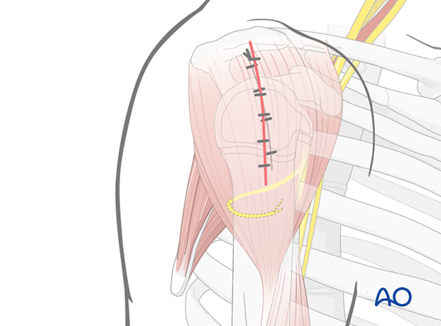
Lateral approach with deltoid splitting
1. Introduction
The transdeltoid lateral approach is an alternative to facilitate reduction of proximal humeral fractures.
This approach provides limited access to the joint, joint capsule, and the biceps tendon.
Usually, there is no need for the adult extensile approach and the approach is limited to management of interposed soft-tissue and fracture reduction.
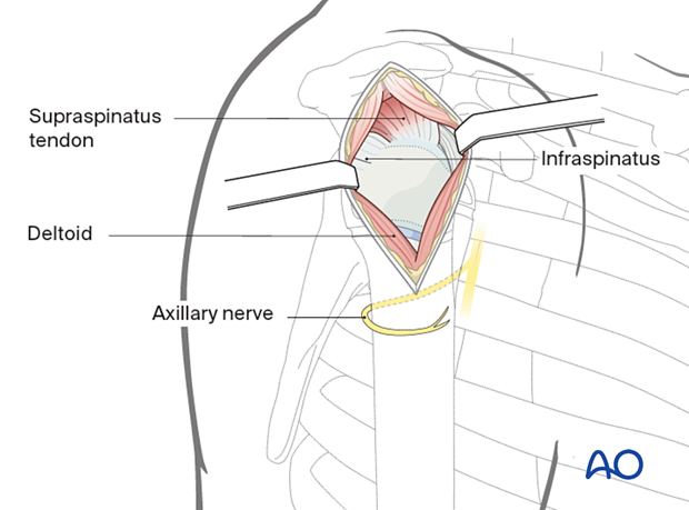
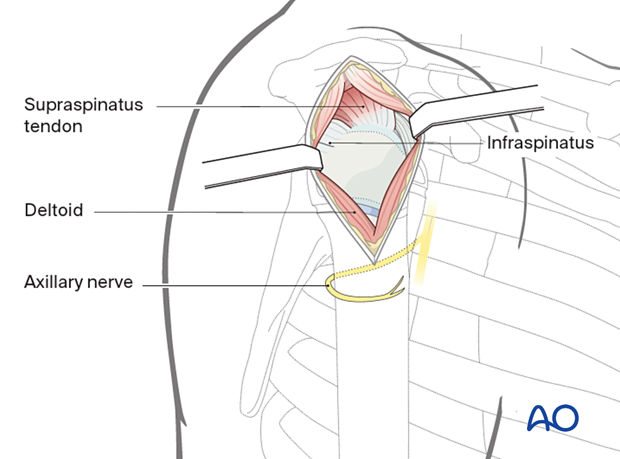
The incision is placed in the middle part of the deltoid muscle, as illustrated.
Depending on the fracture morphology and planned osteosynthesis, the skin incision may be extended distally but the axillary nerve should be protected.
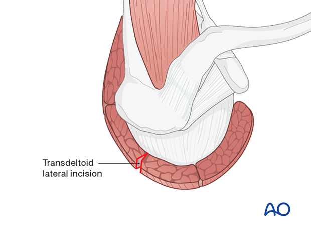
2. Anatomy
Neurovascular structures
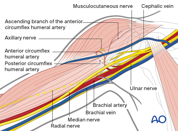
The course of the axillary nerve must be appreciated.
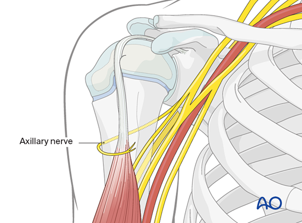
This approach utilizes a relatively avascular plane, away from the anterior and posterior circumflex humeral arteries.
3. Skin incision
Anatomical landmarks
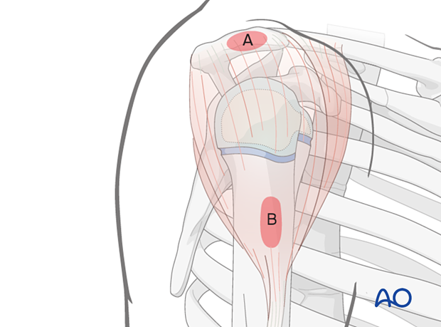
Anatomical landmarks for the transdeltoid lateral approach are:
- Lateral border of the acromion (A)
- Lateral side of the proximal humeral shaft (B)
Both landmarks can easily be palpated.
Axillary nerve
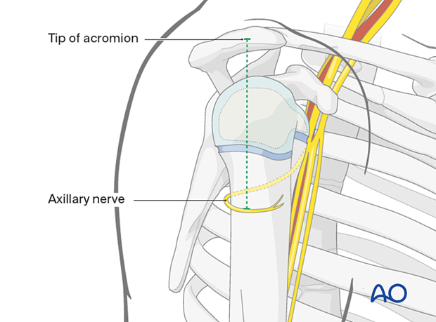
The axillary nerve runs dorsolaterally around the humeral metaphysis on average 5 cm distal to the tip of the acromion in children of 6 years and older.
Skin incision
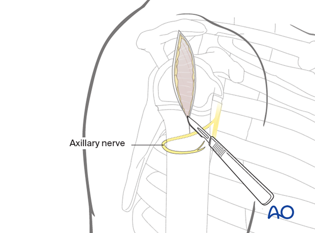
Make a skin incision from the lateral border of the acromion parallel to the axis of the humerus.
4. Exposure of the middle third part of the deltoid muscle
Expose the middle third of the deltoid and split between its fibers.
For maximum exposure, split the deltoid up to the margin of the acromion.
Palpate the axillary nerve on the deep surface of the deltoid muscle, distal to the incision.
Hemorrhagic subdeltoid bursal tissue may require excision to expose the humeral head.
5. Wound closure
Irrigate the wound.
Close the deltoid fascia, subcutaneous tissues, and skin.
