Plate fixation of multifragmentary olecranon fracture
1. General considerations
Pediatric considerations
Bridge plating is used primarily for comminuted olecranon fractures. These fractures often involve a high energy mechanism in brittle metaphyseal bone.
They are rarely seen in pediatric practice and almost always in adolescent patients at or nearing skeletal maturity.
In this case periarticular plates designed for adult patients are often suitable. The residual growth from the olecranon apophysis is minimal and can be ignored.
Children who are too small for such a plate may be treated with a modified 1/3 tubular plate cut and bent into a hook plate configuration.
Every effort should be directed at anatomical reduction and stable fixation of the major articular surface fragments, particularly the coronoid process.
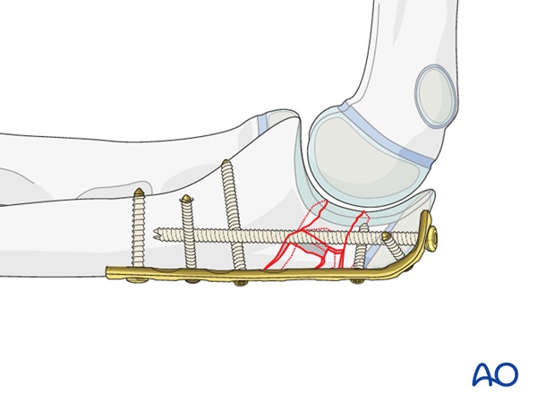
In this situation, the fracture must be bridged without compression to preserve the size and shape of the olecranon fossa. Small interfragmentary screws or K-wires may be used to improve stability of larger fragments.
Bridge plating
In multifragmentary fractures of the olecranon, anatomical reconstruction of the articular surface is the primary goal. Intermediate, nonarticular fragments do not need to be reduced anatomically.
Directly manipulate only the articular fragments. Excessive manipulation of intermediate fragments risks disturbing their blood supply. If the soft-tissue attachments are preserved, and the fragments are relatively well aligned, healing is predictable. Alignment of the main fragments can usually be achieved indirectly using traction and soft-tissue tension.
Mechanical stability, provided by the bridging plate, is adequate for indirect healing (callus formation).
2. Patient preparation
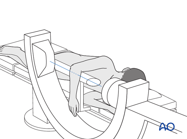
This procedure may be performed with the patient either in a prone position or in a lateral decubitus position.
3. Choice of approach
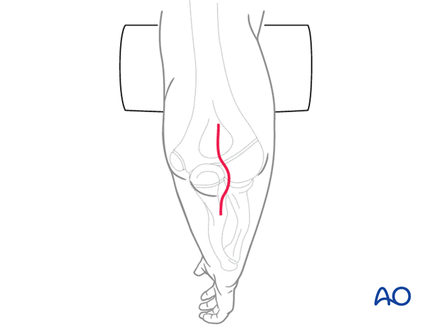
The ulna is exposed through a posterior approach.
An alternative is the posterolateral approach, which provides access to both the proximal ulna and the radial head. A disadvantage of this approach is the potential for cross-union.
4. Reduction
Reduction of articular fragments
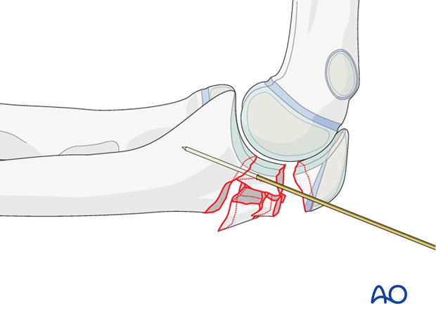
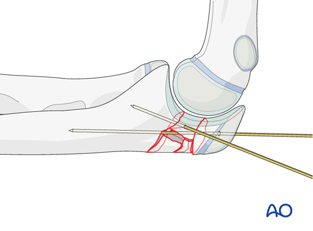
Through a posterior incision, release the joint capsule medially and laterally at the fracture site. If necessary, identify and protect the ulnar nerve. With direct visualization, manipulate and anatomically reduce the articular fragments. Use the distal humerus as a template for reduction. Provisionally fix the fragments with 1.0 mm K-wires.
Reduction of proximal segment
Reduce the proximal part of the olecranon with pointed reduction forceps and temporarily hold the reduction with one or two K-wires.
Insert all K-wires in a position where they do not interfere with the planned plate and screws.
Check reduction with direct visualization of the sigmoid notch and the posterior cortex of the olecranon and confirm with image intensifier.
5. Plate preparation
Implant choice
An adult sized periarticular plate of any design will fit most adolescent elbows. Some of these systems have a left and right side specific to the anatomy. Small diameter 2.7 locking screws with flexible configuration are useful for comminuted fractures.
For smaller children cut a 1/3 tubular plate and fashion it into a hook plate.
Choose the length of the plate so that at least three screws may be inserted in the most proximal fragment and three in the diaphysis.
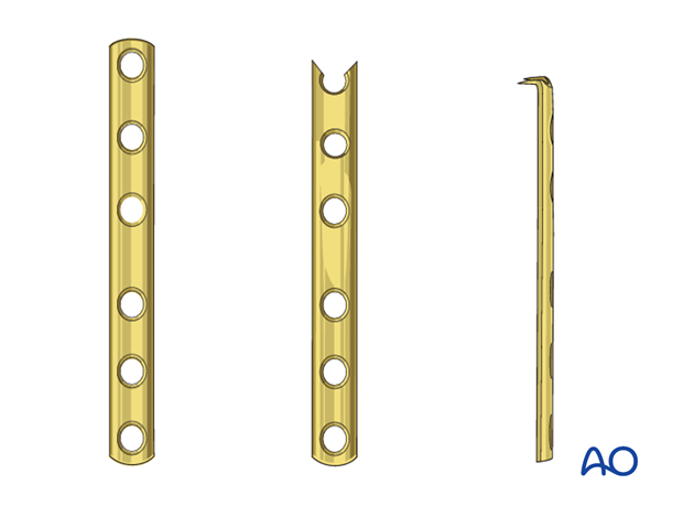
Plate contouring
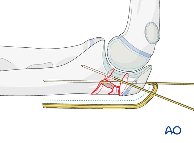
If an anatomic plate is not used, contour the plate to fit the proximal ulna, bending around the tip of the olecranon. Make sure to follow the curvature of the olecranon.
Contouring is done with bending irons and a bending press.
6. Plate fixation
Split of triceps attachment
To achieve close bone plate contact, split the triceps attachment before positioning the plate.
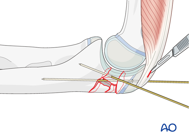
Plate placement and insertion of proximal screws
Hold the plate provisionally to the bone with K-wires and check with an image intensifier before inserting screws.
Anchor the plate with three screws to the proximal olecranon. Make sure that the screws do not protrude into the joint. A long medullary screw can add considerable stability.
Whenever possible, insert the screws bicortically, aiming the drill to the lateral or to the medial cortex.
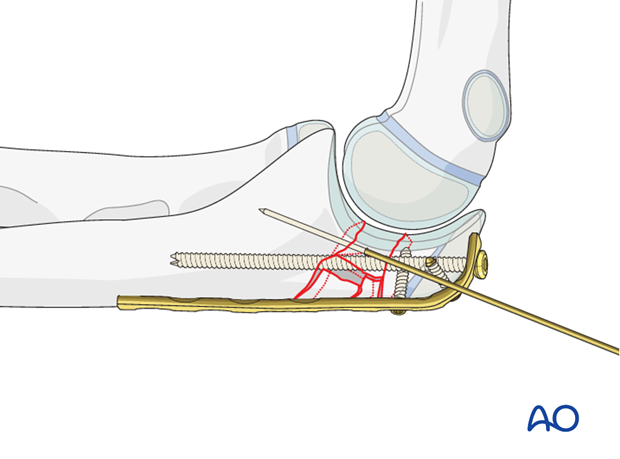
Insertion of distal screws
Insert three divergent bicortical screws into the distal fragment.
Make sure that the contour of the articular surface of the olecranon is maintained
Remove the K-wires.
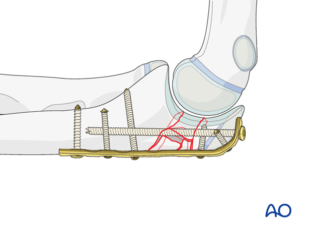
7. Fixation of a coronoid fragment
Reduction of a large coronoid fragment of multifragmentary proximal ulnar fractures may often be performed through the fracture site. Provisional or definitive fixation may be implemented at that time, but sometimes it helps to use a screw through a posterior plate for its fixation, as shown here.
Insert this as a lag screw, as perpendicular as possible to the fracture plane at the base of the coronoid. Smaller diameter screws should be used if possible.
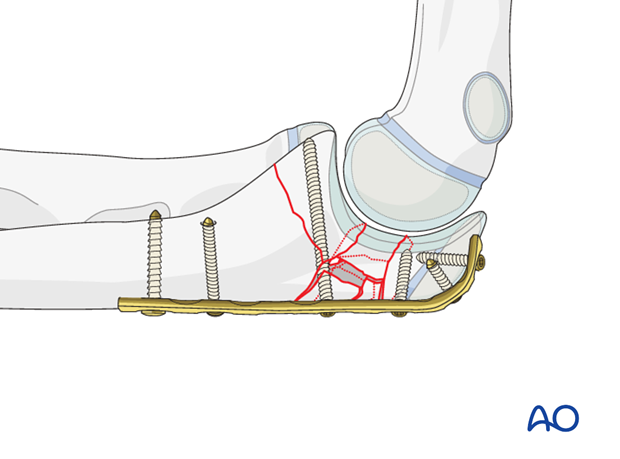
Stabilize a coronoid fragment that is too small for screw fixation with sutures.
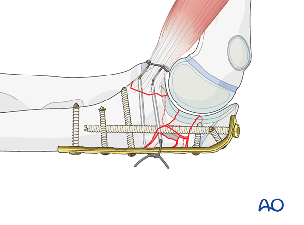
8. Final assessment
Take AP and lateral views.
Confirm that there is no screw penetration into the joint.
Examine for full pronation/supination and for construct stability through flexion/extension.
