Open reduction, tension band (greater trochanter)
1. Preliminary remarks
Introduction
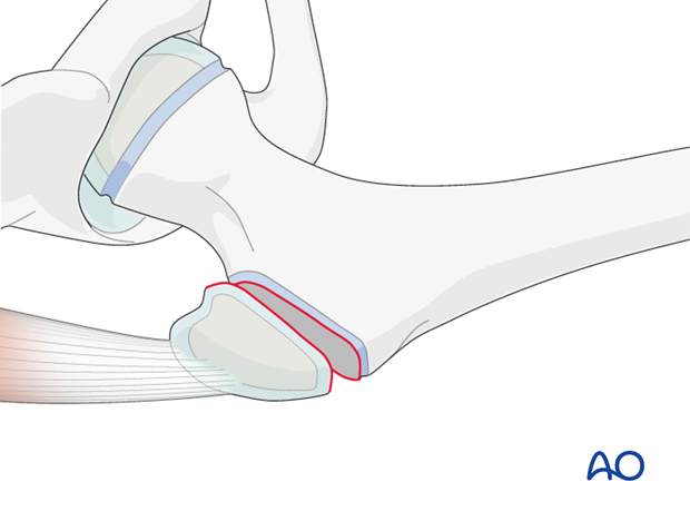
Avulsion of the greater trochanter that disrupts the abductor mechanism can be repaired by open reduction and screw or tension band fixation depending on the size of the avulsed fragment.
A tension band wire technique is a good alternative for smaller fragments.
For children with 4 or more years of growth remaining, it is recommended that implants are removed after fracture healing to allow for growth.
For children with less than 4 years of growth remaining, the implants can be left in place unless symptomatic.
Equipment selection
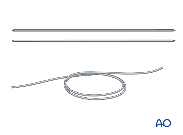
The following implants are required:
- 1.6, 2.0, or 2.5 mm K-wires
- 1.0, 1.2, or 1.4 mm cerclage wire or heavy suture material
2. Patient preparation and approach
Patient preparation
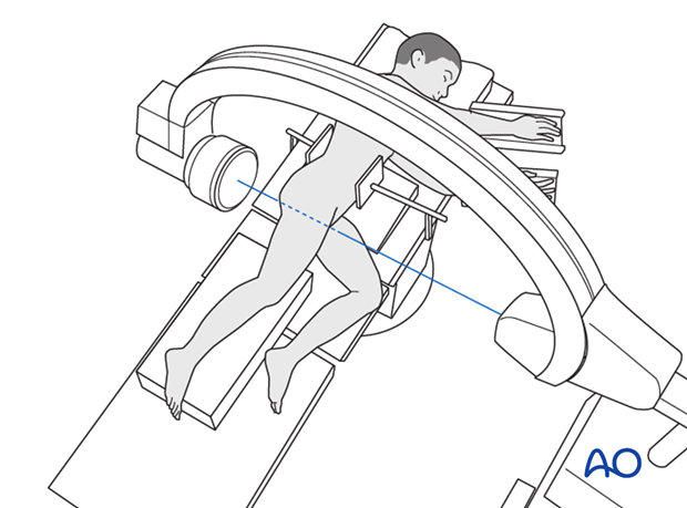
This procedure is normally performed with the patient in a lateral position.
Approach
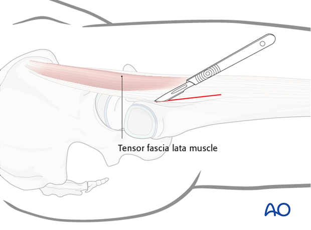
The fracture fragment is approached with a lateral approach, the hematoma is evacuated and periosteum is cleared to allow accurate reduction of the fragment(s).
3. Reduction
The trochanteric fragment is directly reduced to its bed and the position confirmed using image intensification.
A towel clip may be used for temporary control, or a K-wire can be used to aid manipulation and to provide temporary fixation.
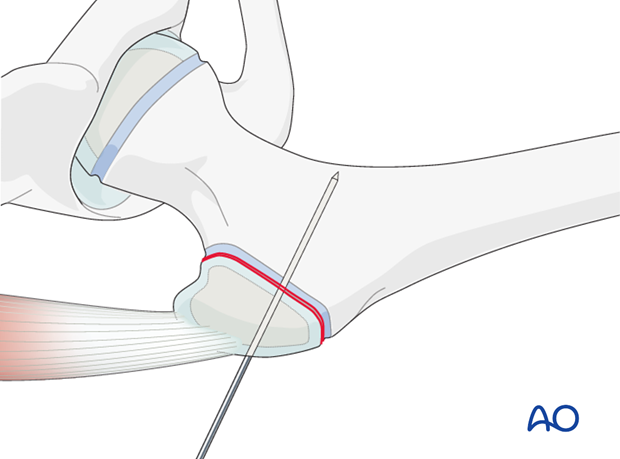
4. Fixation
K-wire placement
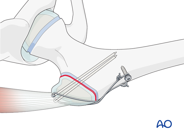
Two K-wires are placed superiorly in the trochanter. The trajectory should be perpendicular to the fracture line with one wire slightly anterior and the other slightly posterior. Tips of the wires should just perforate the dense bone of the medial calcar, if possible. The wires may be backed out to allow for later impaction.
If a temporary K-wire has been used, it can now be removed.
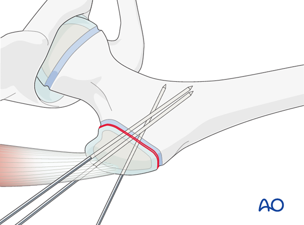
Preparation for wire insertion
An anterior to posterior hole is drilled through the lateral metaphysis of the proximal femur using a 2.0 or 2.5 mm drill bit.
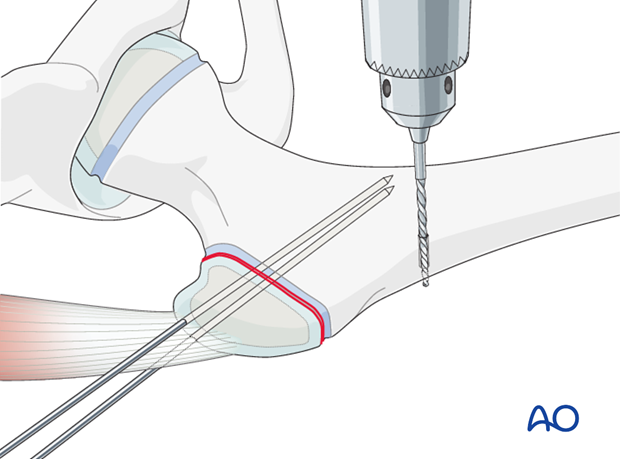
Wire preparation and insertion
The cerclage wire is prepared by making a loop approximately one third along its length.
The shorter segment of the wire is inserted through the drill hole.
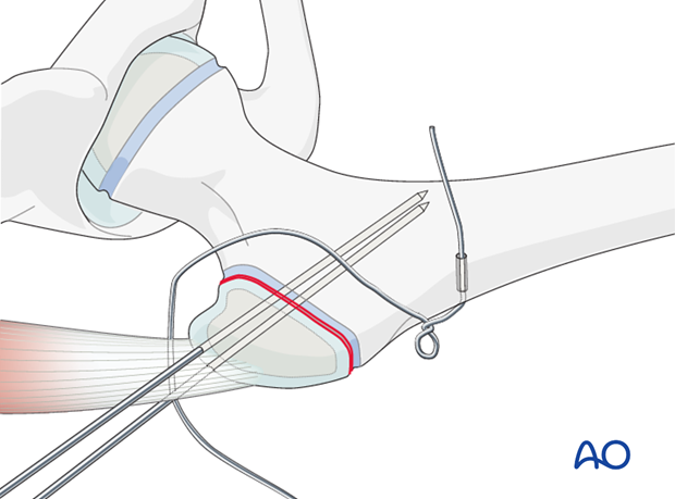
The long segment of the wire (bearing the loop) is passed in a figure-of-eight configuration, deep to the abductor tendon around the protruding ends of the K-wires.
Pearl: Passing this K-wire can be facilitated by initially inserting a 14-gauge Angiocath in the desired position, removing the metal needle, and passing the cerclage wire though the plastic cannula.
The cerclage wire ends are united with a little twist.
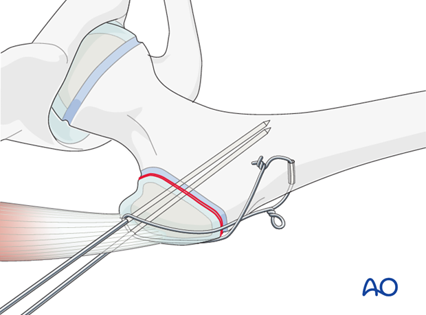
The wire ends are cut short.
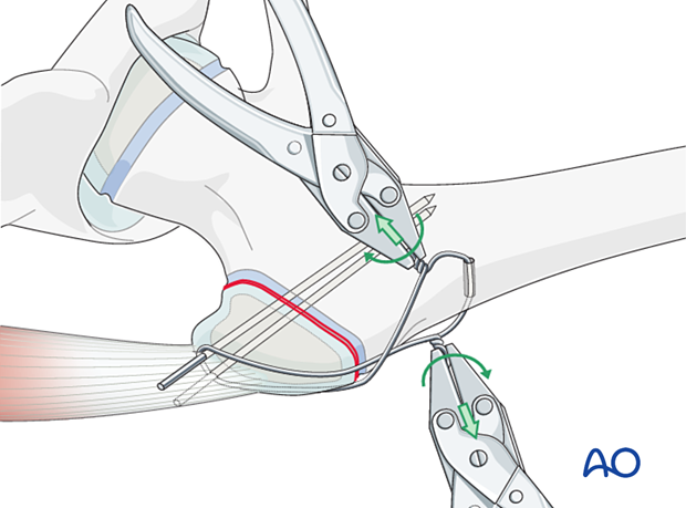
The slack is then taken up by further twisting. This is repeated until the desired tension is achieved. Both loops must be tightened at the same time and in the same direction, in order to achieve equal tension on both arms of the wire.
By simultaneously tightening the twist and the loop with two pliers, the two fragments are drawn together such that the fracture is placed under compression.
The twisted wire is trimmed and both ends turned towards the femur in order to prevent subsequent irritation of the soft tissues.
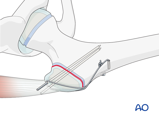
Sinking the K-wires
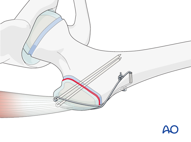
A plier, bending iron, and forceps are used to bend the K-wires through 180°.
The K-wires are then driven home, sinking their curved ends into the bone in order to prevent backing out.
Alternative: Cortical screw anchor
A 4.5 mm cortical screw can be used to anchor the cerclage wire distally.
Alternative: Nonresorbable suture
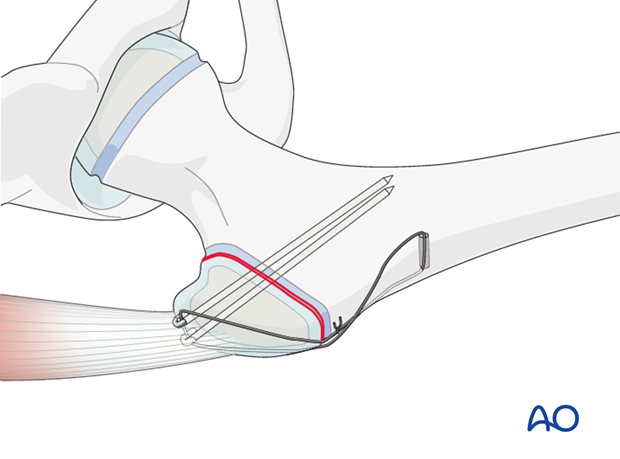
A nonresorbable suture can be used as a tension band instead of wires. This may be more suitable in younger patients.
Closure
Routine closure according to the surgeon's preference.
5. Aftercare
Introduction
Range-of-movement exercises should start in the immediate postoperative period to prevent stiffness. Surgeons should indicate if any extremes of movement should be avoided.
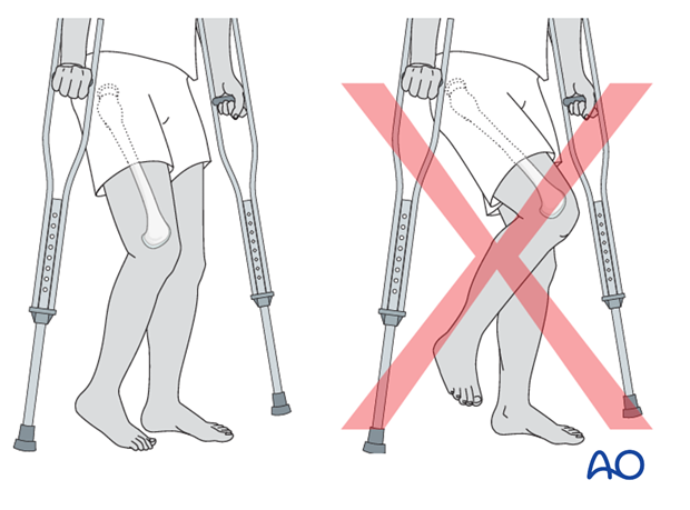
Forces through the hip are less with toe-touch weight bearing than with non-weight bearing.
Crutch walking with toe-touch weight bearing should therefore be advised for 3–4 weeks.
This needs to be taught to children and supervised by a physiotherapist.
Abductor strengthening exercises can be started after 6–8 weeks if there are clinical and radiological signs of healing.
Infection
See the additional material on postoperative infection.
Weight bearing
Having started with toe-touch weight bearing, children progress to partial weight bearing and then to full weight bearing according to their age and the predicted rate of healing of their fracture.
Even older adolescents should be fully weight bearing without aids at three months.
Sports
Swimming can be allowed as soon as partial weight bearing is permitted.
Contact sports should be avoided for at least six months.
Follow-up x-rays
X-rays are generally taken immediately after the surgery and at 6 and 12 weeks.
Implant removal
The fracture should be healed and consolidated prior to implant removal (see Healing times).
Implants in young children should always be removed to prevent them from being covered by bony overgrowth.
Implant removal is not compulsory in adolescents.


