

Osteosynthesis in pediatric polytrauma
1. Introduction
In pediatric polytrauma, the timing and method of fracture stabilization are determined not only by the fracture pattern but also by the patient’s physiological condition.
Initial management prioritizes life-saving interventions and restoration of hemodynamic stability. In unstable patients, rapid preliminary fracture stabilization using damage control principles is often preferred to minimize the additional physiological burden of surgery.
Definitive osteosynthesis can be undertaken primarily in selected stable patients or secondarily once resuscitation is complete and the patient’s clinical and soft-tissue conditions are favorable.
Whereas in adults, the fracture fixation philosophy has moved more to “damage control orthopedics” and away from total immediate surgical care, there are very few studies on the optimal timing of fracture fixation in children. Valid studies are needed in this field to facilitate evidence-based decision-making about the timing of fracture stabilization and definitive osteosynthesis in polytraumatized children.
For management of specific fractures, refer to the corresponding anatomical module in the pediatric trauma section in AO Surgery Reference.
2. External fixation
Frequently, external fixation is performed primarily as damage control during early resuscitation and patient stabilization.
External fixation is used for preliminary stabilization of a limb.
It may also be used as definitive fixation of the fracture in selected cases, eg, in femoral and tibial fractures, under the following conditions:
- No bone loss
- Good alignment achieved
- Stable fixation achieved
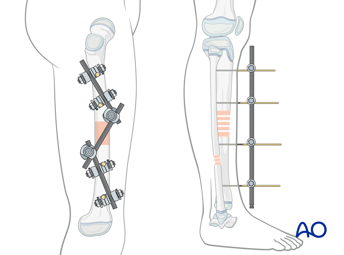
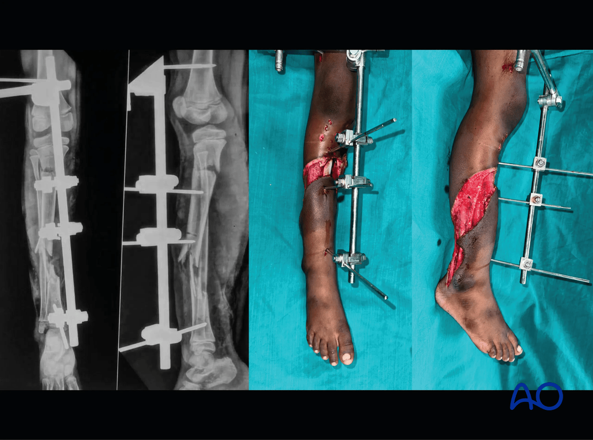
Definitive management of an open segmental tibia shaft fracture with external fixation in an 8-year-old patient.
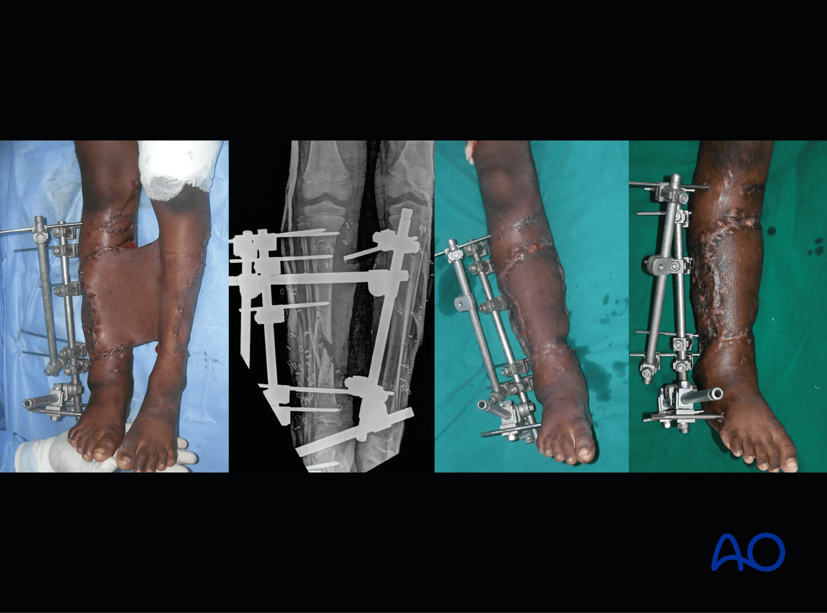
X-ray and clinical pictures after cross-leg flap. Note that the external fixator is realigned sparing the knee joint to allow knee mobilization after flap healing.
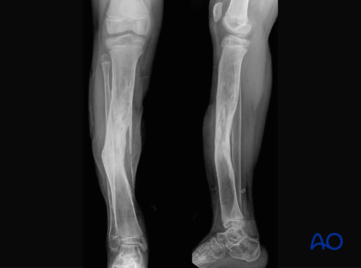
Four years follow-up x-ray showing good healing of the fracture despite the initial bone loss at the distal fracture site without any additional intervention. This shows the power of the periosteum in children to heal fractures.
3. Internal fixation
Primary fracture stabilization
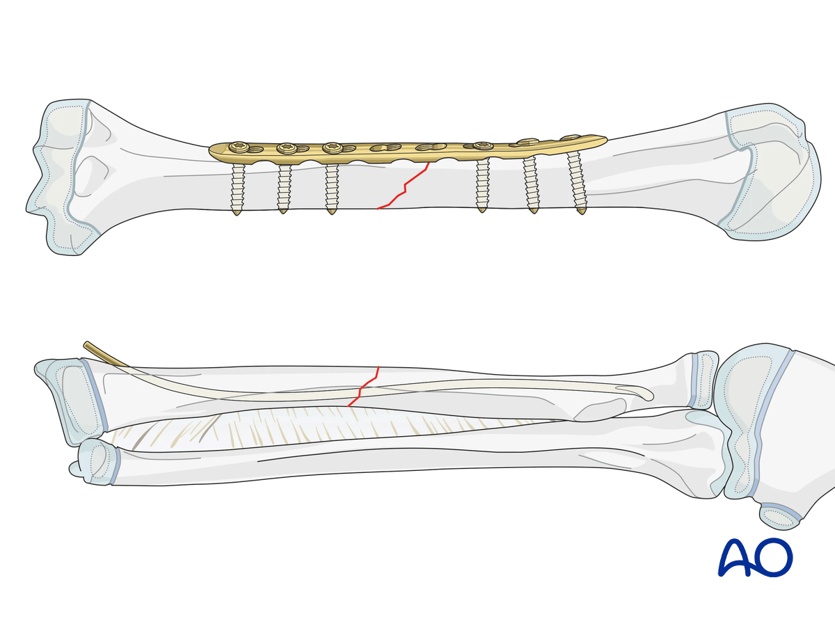
In selected cases, internal fixation may be opted for primary stabilization of the fractures. This is more suitable in the upper limb where good muscular and vascular coverage has a lower risk of infection.
Examples of anatomical areas with internal fixation:
- Humeral fractures
- Both bone fractures of the forearm
Prerequisites for internal fixation:
- Good wound debridement, no contamination
- Early patient stabilization possible
- Favorable (eg, simple) fracture configuration for internal fixation
Secondary fracture stabilization
Once the patient is hemodynamically stable, conversion of a preliminary external fixation to internal fixation may be considered.
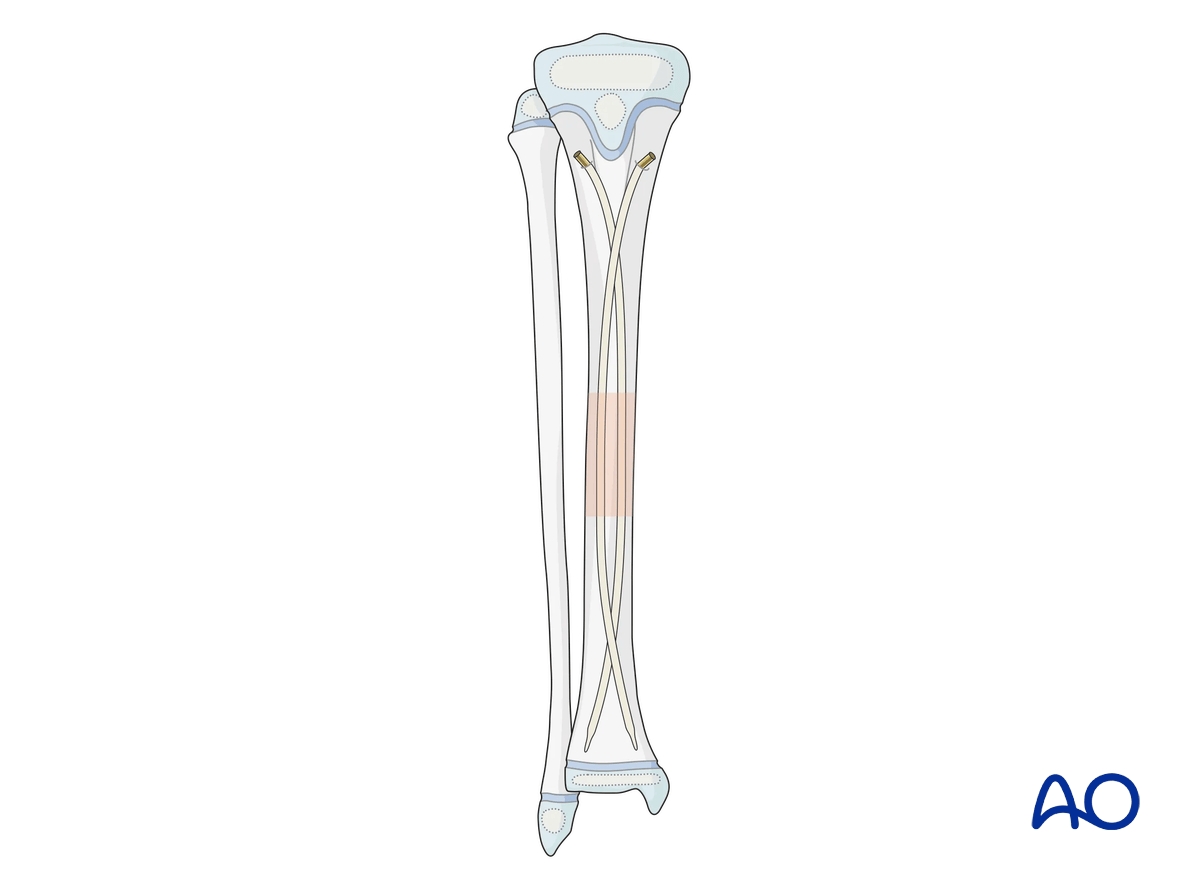
The soft-tissue status needs to be evaluated. Polytrauma patients experience high-energy trauma with open fractures. The wound needs to be healthy without any signs of infection.
The internal fixation surgery may be combined with soft-tissue coverage (flap/skin grafting).
Management of bone loss
If there is bone loss (<4 cm), the bone may be acutely shortened and stabilized in alignment. A bone elongation can then be performed at a later stage. This approach also helps to manage any soft-tissue compromise.
For patients with larger bone loss (>4 cm), bone transport has to be planned after adequate soft-tissue healing.
Management of complications and sequelae
A pediatric patient with an open fracture can have delayed or nonunion unlike closed fractures and may need secondary interventions to get the fracture to heal completely. A nonunion following an open fracture will need either bone-grafting or revision osteosynthesis with bone grafting.
An infected nonunion (FRI) is another potential complication in a polytraumatized child with a contaminated open fracture. These patients will need radical debridement for infection control followed by osteosynthesis or bone transport depending on the bone gap after debridement.
The third potential complication is growth arrest leading to secondary deformities or limb shortening in children with physeal injuries (injury to the growth plate). It is pertinent to watch out for these problems during follow-up and deal with them appropriately.
4. Reference
- Nahm NJ, Como JJ, Wilber JH, et al. Early appropriate care: Definitive stabilization of femoral fractures within 24 hours of injury is safe in most patients with multiple injuries. J Trauma. 2011 Jul;71(1):175–185.


