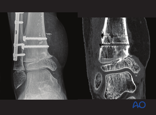
Open reduction; screw fixation
1. General considerations
Introduction
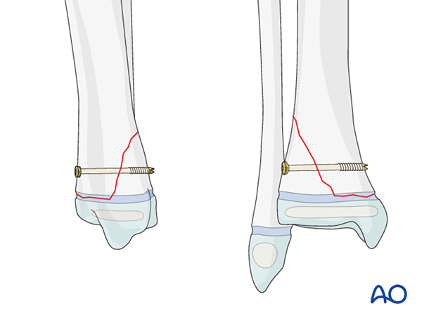
A Salter-Harris II fracture may be fixed with a lag screw through the metaphyseal (Thurstan Holland) fragment and the metaphysis of the proximal fragment, parallel to the physis.
If the metaphyseal fragment is large enough, two screws may be used.

The metaphyseal fragment is either posterior, posterolateral, or lateral.
If the metaphyseal fragment can be reduced easily, the screws can be inserted directly on the side of the metaphyseal fragment.
For a posterior Thurstan Holland pattern, the screws are typically inserted from anterior to posterior.
For a lateral fragment, the screws may be placed either from posteromedial or lateral anterior to the fibula.
If there is medial soft-tissue damage, lateral screw insertion is recommended.
Associated fibular fracture
A fibular fracture often reduces with reduction and fixation of the tibial fracture and does not require separate consideration.
If the alignment and stability of the fibular fracture are unsatisfactory after fixation of the tibial fracture, surgical treatment of the fibular fracture is also required.
If the distal tibial fracture is highly comminuted, fixation of the fibular fracture may add to overall stability.
Treatment goals
The main treatment goals are:
- Stabilize the fracture
- Minimize further physeal injury
Physeal fractures may be complicated by growth disturbance. To minimize the risk, reduction should be anatomical and conducted with minimal force.
Closed vs open reduction
If initial closed reduction is unsuccessful, this is usually due to periosteum entrapped in the fracture on the side that has failed in tension.
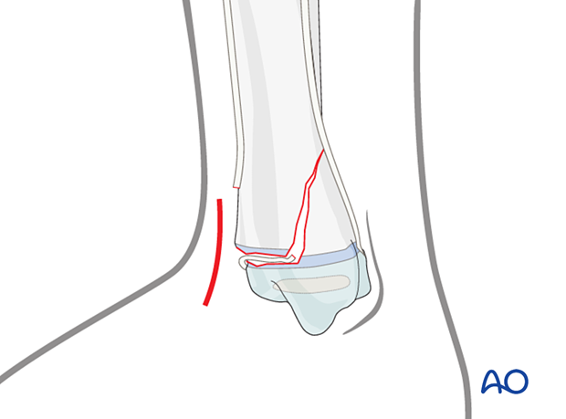
The initial incision is made on the side opposite to the metaphyseal fragment that remains attached to the epiphysis.
2. Instruments and implants
Appropriately sized cannulated or noncannulated lag screws (2.7, 3.5, or 4.0 mm) can be used.
The following equipment is used:
- Cannulated screw set with guide wire
- Drill
- Image intensifier

3. Patient preparation and approaches
Patient positioning
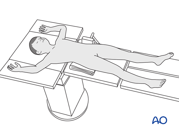
Place the patient in a supine position on a radiolucent table.
Approaches
The approach is dictated by the fracture pattern and location of the block to reduction (eg, periosteum) and includes:
The incision should provide sufficient exposure to allow removal of any block to reduction.
4. Reduction
Remove blood clots, loose fragments, soft callus, and entrapped periosteum.
Reduce the fracture with gentle manipulation.
A hook or reduction forceps may be used.
Confirm reduction with an image intensifier.
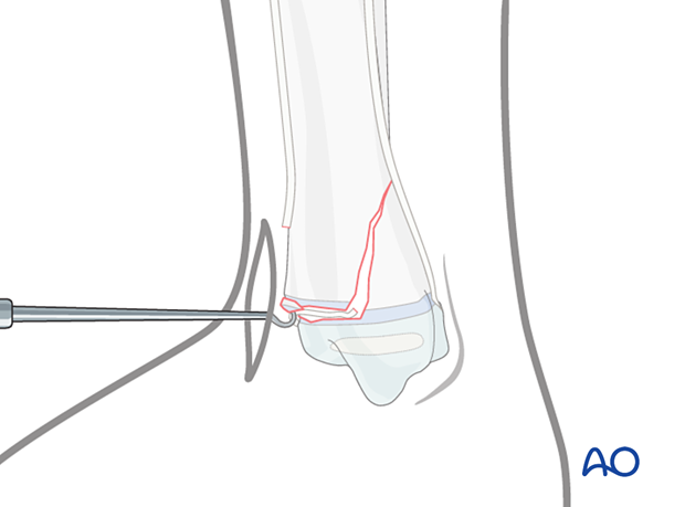
If needed, temporarily stabilize the fracture with K-wire(s) or with pointed reduction forceps.
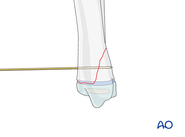
5. Fixation
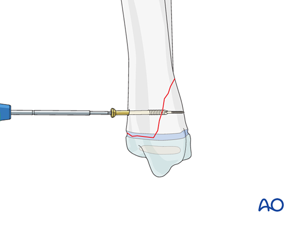
Insert a lag screw in the chosen direction, aiming for the center of the metaphyseal fragment, in a standard manner.
If the metaphyseal metaphyseal fragment is sufficiently large, insert two screws.
Confirm reduction, fracture stability, and screw placement with an image intensifier.
6. Fibular fracture management
Most fibular fractures do not require treatment. Indications for fixation include:
- Augmentation of the stability of tibial fracture fixation
- Significant displacement of the fibular fracture
The type of fracture pattern dictates the method of fixation of the fibular fracture.
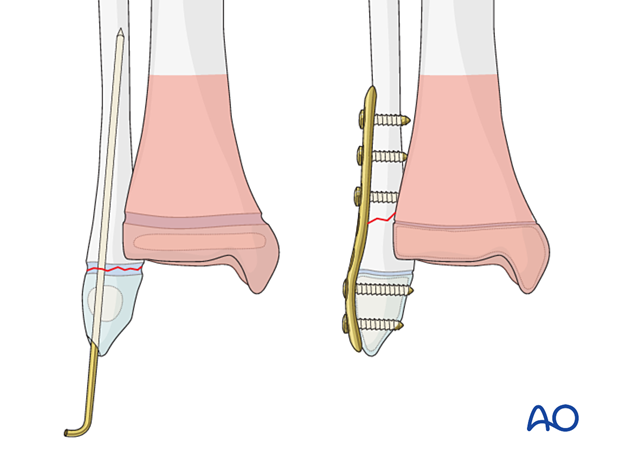
In a younger child, these fractures may be fixed with K-wires in a standard manner. Multiple passes of the K-wire through the physis should be avoided.
In an older patient with a closing physis, these fractures may require plate fixation.
If screws are inserted on both sides of the physis, compression should be avoided and the periosteum and perichondral ring not be disturbed. To protect the perichondral ring, a dissector or elevator may be used to offset the plate during screw insertion. The plate should be removed soon after the fracture has healed.
7. Final assessment
Recheck the fracture alignment and implant position clinically and with an image intensifier before anesthesia is reversed.
Confirm stability of the fixation by moving the ankle through a range of dorsi/plantar flexion.
8. Immobilization
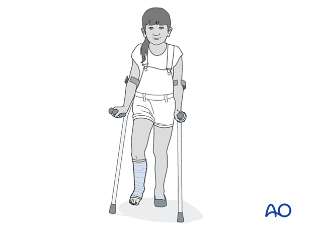
A molded below-knee cast or fixed ankle boot is recommended for a period of 2–6 weeks as the strength of fixation may not provide sufficient stability for unrestricted weight bearing.
9. Aftercare
General considerations
Weight-bearing status for these fractures is based on local protocol and should consider the severity of soft-tissue injury and stability of fixation.

Pain control
Patients tend to be more comfortable if the limb is splinted.
Routine pain medication is prescribed for 3–5 days after surgery.
Neurovascular examination
The patient should be examined frequently to exclude neurovascular compromise or evolving compartment syndrome.
Discharge care
Discharge follows local practice and is usually possible within 48 hours.
Follow-up
The first clinical and radiological follow-up is usually undertaken 5–7 days after surgery to check the wound and confirm that reduction has been maintained.
Cast removal
A cast or boot can be removed 2–6 weeks after injury.
Mobilization
After cast removal, graduated weight-bearing is usually possible.
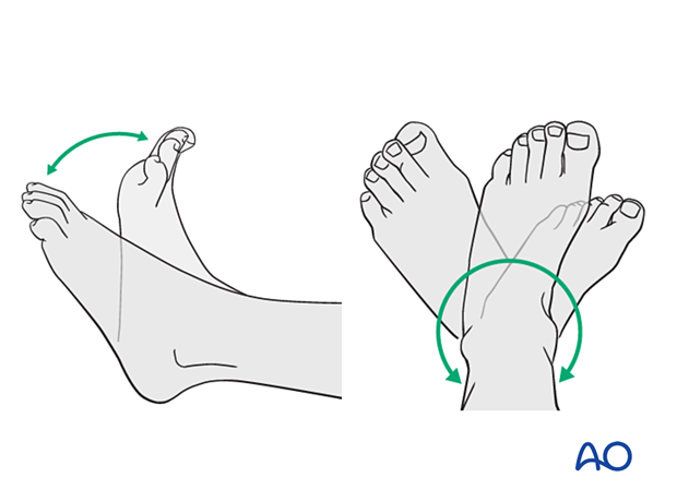
Patients are encouraged to start range-of-motion exercises. Physiotherapy supervision may be required in some cases but is not mandatory.
Sports and activities that involve running and jumping are not recommended until full recovery of local symptoms.
Implant removal
Implant removal is not mandatory and requires a risk-benefit discussion with patient and carers.
Follow-up for growth disturbance
All patients with distal tibial physeal fractures should have continued clinical and radiological follow-up to identify signs of growth disturbance.
Compare alignment and length clinically with the uninjured leg.
A Harris growth arrest line, parallel to the physis, is radiological evidence of continuation of normal growth.
Recommended reading:
- Nguyen JC, Markhardt BK, Merrow AC, et al. Imaging of Pediatric Growth Plate Disturbances. Radiographics. 2017 Oct;37(6):1791–1812.
Parallel Harris growth arrest line in the distal tibia of a 10-year-old patient, 6 months after injury (right image) excluding a physeal injury
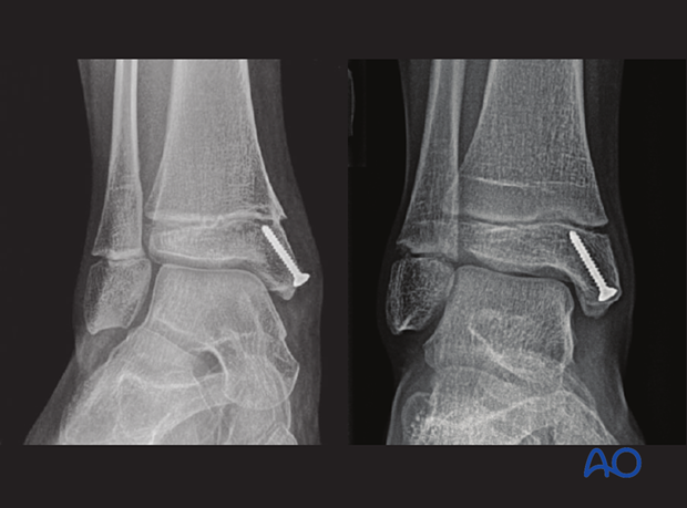
Growth disturbance (convergent Harris growth arrest lines) of an open Salter-Harris II fracture of the distal tibia in a 10-year-old patient, 6 months after injury
