Posterolateral approach to the pediatric distal tibia
1. General considerations
The posterolateral approach gives direct access to posterior and posterolateral tibial fragments. It may also be used to access the posterior aspect of the distal fibula.
For this approach, the patient is positioned in a prone or lateral position.
2. Skin incision
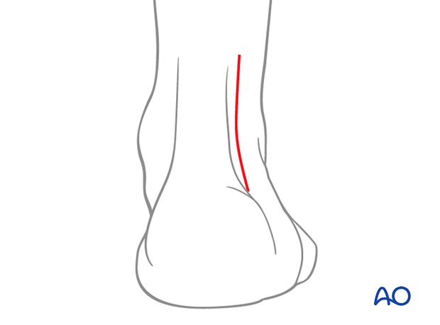
Make an incision 0.5 cm lateral and parallel to the lateral border of the Achilles tendon.
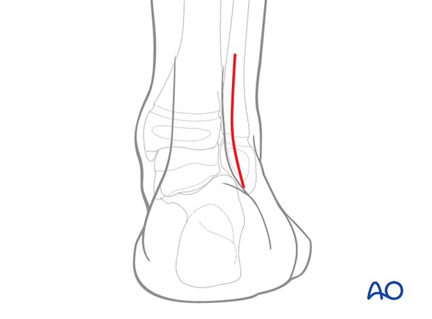
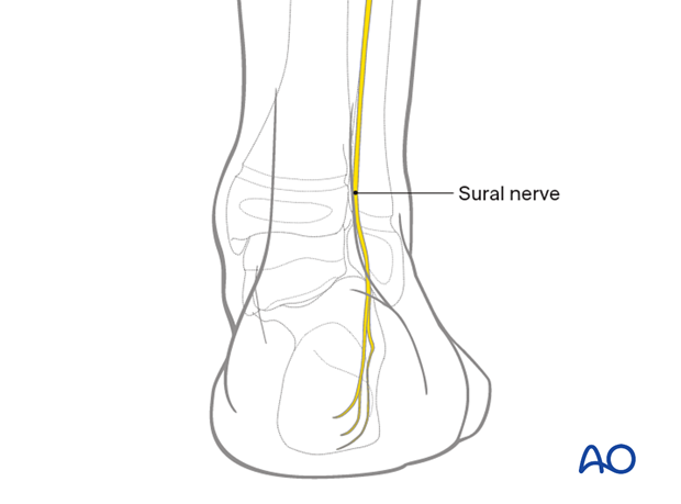
The sural nerve must be protected. If the dissection is extended more proximally, it may be necessary to work on either side of this nerve.
Dissect through the skin and subcutaneous tissues to expose the peroneal muscles.
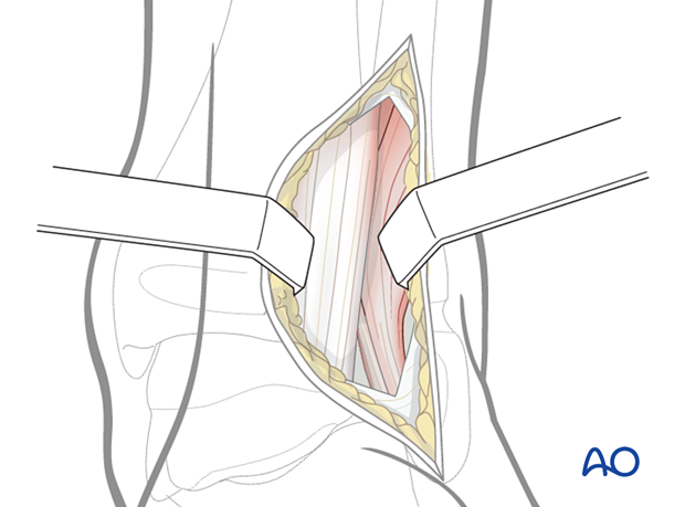
Dissect the medial border of the peroneal muscles and retract them laterally.
This will expose the posterior and posterolateral tibial fragments.
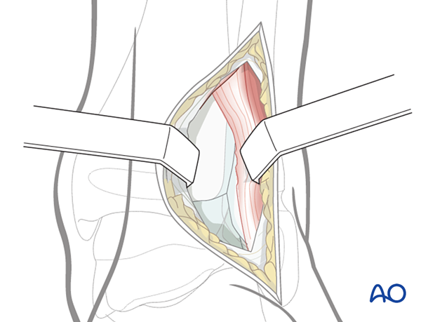
3. Wound closure
At the end of the procedure, the peroneal muscles are returned to their anatomical position.
The deep fascia may be closed over the peroneal muscles if this can be achieved without tension.
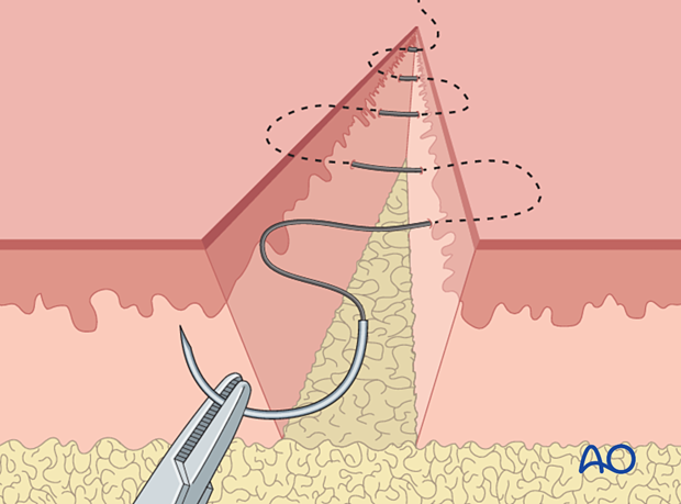
After careful hemostasis, close the subcutaneous tissue and skin separately. Subcuticular, absorbable skin sutures may be used if the condition of the soft tissues permits it.
