Excision
1. General considerations
Reconstruction vs excision
Reasonable attempts should be made to repair the articular surface, but there may be small osteochondral defects that are not reconstructable.
A small, loose osteochondral fragment in the ankle may cause more symptoms than a small articular defect.
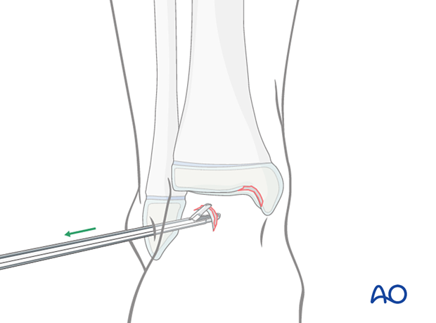
Small articular fragments that are not amenable to reconstruction may be removed arthroscopically or with an open approach.

2. Patient preparation and approaches
Patient positioning
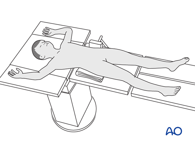
Place the patient in a supine position on a radiolucent table.

Approaches
When appropriate resources are available, arthroscopically assisted excision is recommended.
If arthroscopy is not available, small osteochondral fragments can be excised through an open approach that depends on the location of the fracture.

3. Excision
Fragment removal assisted by arthroscopy
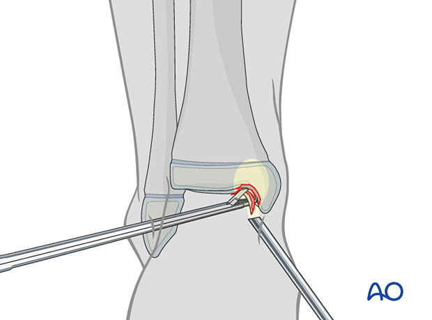
Identify and grasp the fragment under arthroscopic control.

4. Final assessment
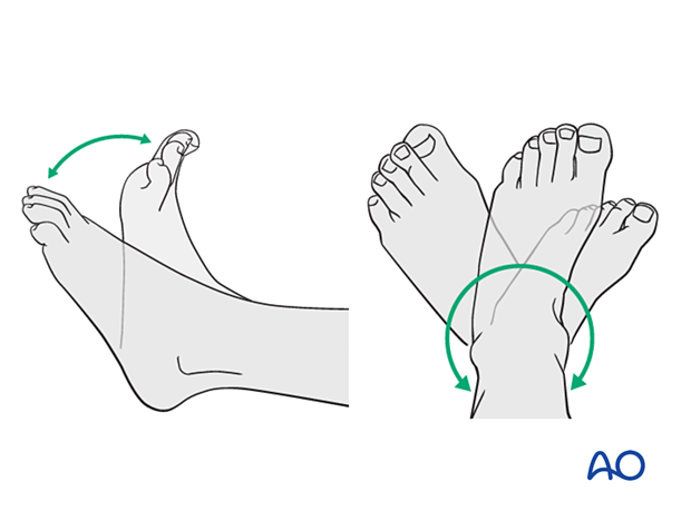
Carefully examine the range of motion and ligament stability.
5. Aftercare
Immediate postoperative care
The patient should get out of bed and begin ambulation with crutches on the day of surgery or the first postoperative day.
In most cases, the patient is mobilized without a cast.
Pain control
Routine pain medication is prescribed for 3–5 days after surgery.
Neurovascular examination
The patient should be examined frequently to exclude neurovascular compromise or evolving compartment syndrome.
Discharge care
Discharge follows local practice and is usually possible within 48 hours.
Mobilization
Graduated weight-bearing is usually possible.
Patients are encouraged to start range-of-motion exercises. Physiotherapy supervision may be required in some cases but is not mandatory.
Sports and activities that involve running and jumping are not recommended until full recovery of local symptoms.

Follow-up
The first follow-up is usually undertaken 5–7 days after surgery to check the wound.


