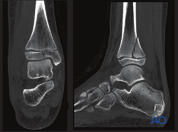
Highlights of Pediatric distal tibial and fibular fractures
Introduction
Publication of first edition on September 30, 2022
The content of the pediatric distal tibial and fibular fracture management has been authored by Daniel Green (US), Philip Henman (UK), and Mamoun Kremli (SA) with Fergal Monsell (UK) and James Hunter (UK) as editors.
Visit the module entry page here!

Highlights
This module covers the treatment options for:
- Metaphyseal fractures of the distal tibia and fibula
- Epiphyseal fractures of the distal tibia and fibula
- Injuries to the syndesmosis
Besides the Salter-Harris fractures, it describes in detail the Tillaux and triplane fractures and their management.
Recommendations by the editors
The section on triplane fractures contains a logical approach to fixation. It is accompanied by a description page with very clear illustrations of the fracture pattern in three dimensions.
Before attending a pediatric fracture course, it is recommended to read about Salter-Harris III and IV, and triplane fractures, as these frequently cause difficulty to delegates.
Content
Table of contents
The pediatric material of the distal tibia and fibula, first edition, includes a detailed description of the following fracture types and their management.
Metaphyseal tibia
Torus/buckle fracture:
- Short leg cast
Simple complete fracture:
- Short leg cast
- Closed reduction; short leg cast
- Closed reduction; K-wire fixation
- External fixation
- Elastic nailing
- MIO bridge plating
Multifragmentary complete fracture:
- Long leg cast
- Closed reduction; long leg cast
- Closed reduction; K-wire fixation
- External fixation
- Elastic nailing
- MIO bridge plating
- Open reduction; plate fixation
Metaphyseal fibula
Torus/buckle fracture:
- Short leg cast
Complete fractures:
- Short leg cast
- Closed reduction; short leg cast
- Open reduction; plate fixation
Epiphyseal tibia
Epiphysiolysis (Salter-Harris I):
- Short leg cast
- Closed reduction; short leg cast
- Closed reduction; K-wire fixation
- External fixation
- Open reduction; K-wire fixation
Epiphysiolysis with metaphyseal wedge (Salter-Harris II):
- Short leg cast
- Long leg cast
- Closed reduction; short leg cast
- Closed reduction; long leg cast
- Closed reduction; K-wire fixation
- External fixation
- Closed reduction; screw fixation
- Open reduction; K-wire fixation
- Open reduction; screw fixation
Epiphyseal fractures (Salter-Harris III):
- Long leg cast
- External fixation
- Open reduction; K-wire fixation
- Open reduction; screw fixation
Epi-/metaphyseal fractures (Salter-Harris IV):
- Long leg cast
- External fixation
- Open reduction; K-wire fixation
- Open reduction; screw fixation
- Open reduction; plate fixation
Anterolateral epiphyseal fractures (Tillaux fractures):
- Long leg cast
- Open reduction; screw fixation
Complex epi-/metaphyseal fractures (triplane fractures):
- Long leg cast
- External fixation
- Open reduction; screw fixation
Avulsion fractures of the medial malleolus:
- Short leg cast
- External fixation
- Open reduction; screw fixation
Osteochondral fractures (intraarticular flake):
- Short leg cast
- Open reduction; pin or screw fixation
- Excision
Epiphyseal fibula
Epiphysiolysis (Salter-Harris I and II):
- Short leg cast
- Closed reduction; short leg cast
- Closed reduction; K-wire fixation
- Closed reduction; screw fixation
- Open reduction; K-wire fixation
- Open reduction; screw fixation
- Open reduction; plate fixation
Malleolar/ligament injuries
Syndesmotic injuries:
- Short leg cast
- Stabilization with screws
- Stabilization with suture and button (suspensory fixation)
Additional material
The following additional material is available for this module.
Approaches:
- Anteromedial approach to the pediatric distal tibia
- Anterolateral approach to the pediatric distal tibia
- Lateral approach to the pediatric distal fibula
- Posterolateral approach to the pediatric distal tibia
- Minimally invasive approach to the pediatric distal tibia
- Entry points for antegrade elastic nailing in the distal tibia
- Safe zones for pin placement in the tibia and foot
Patient preparations:
- Supine position
- Lateral decubitus position
Further reading:
- Clinical evaluation
- Radiological evaluation
- Comlications and technical failures


