43t-E/6 Complex epi-/metaphyseal fractures of the distal tibia (triplane)
Introduction
The triplane fracture of the distal tibia extends through the physis into the epiphysis and metaphysis. Typically, the metaphyseal and epiphyseal components are in different planes and are connected by the physeal fracture.
Triplane fractures are classified as 43t-E/6.
A two-part fracture (43t-E/6.1) is more common, but there may be additional fragmentation at the level of the epiphysis resulting in a three- or four-part fracture (43t-E/6.2).
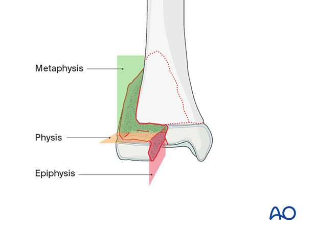
This injury is usually a result of an external rotation force on the plantarflexed foot and ankle.
These fractures occur in adolescents approaching skeletal maturity, when the tibial physis is closed on the medial side. The open lateral component of the distal tibial physis fails with a fracture line that involves the metaphysis and epiphysis.
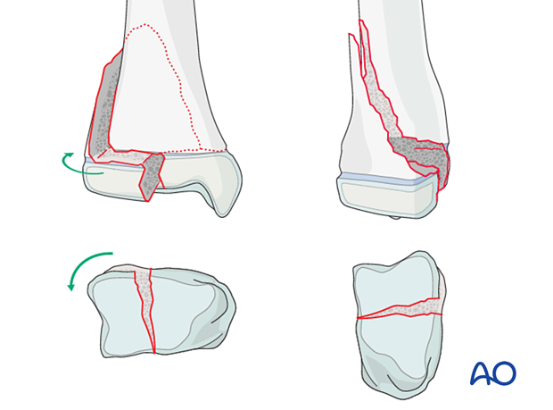
Two-part triplane fracture
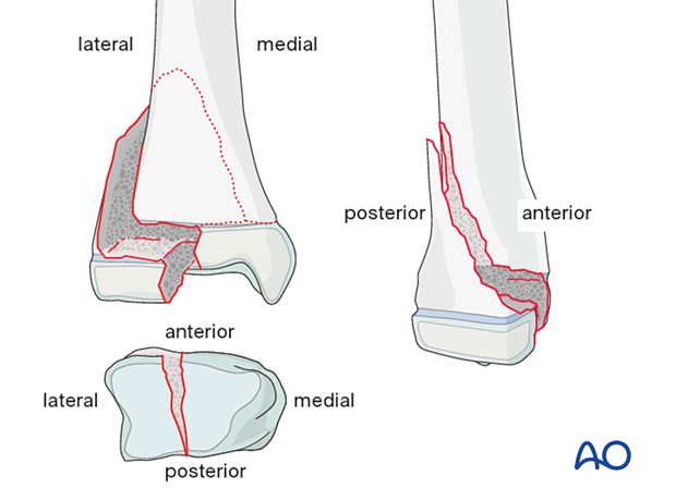
The fracture line in a two-part fracture involves the sagittal, axial, and coronal planes.
There is a fracture through the epiphysis, a horizontal fracture through the physis, and an oblique fracture through the posterolateral metaphysis.
- The lateral epiphysis is connected to the posterior metaphyseal fragment
- The anteromedial epiphysis remains attached to the distal tibia
Imaging
The fracture appears as a Salter-Harris II fracture on a lateral x-ray and a Salter-Harris III fracture on an AP or mortise x-ray.
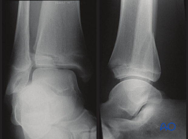
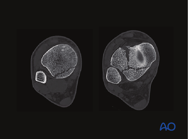
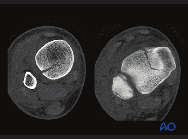
A CT scan is recommended to measure articular displacement and identify the fracture pattern to determine screw trajectory.
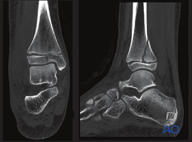
On the left, an axial cut of the metaphyseal component of the fracture is shown; on the right, the cut is at the level of the epiphysis.
Three- and four-part triplane fractures
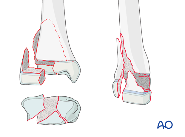
Three- and four-part fractures are associated with additional fragmentation at the level of the epiphysis.
A three-part fracture involves:
- The anterolateral epiphysis
- The posterior epiphysis, connected to the posterior metaphyseal fragment
- The medial epiphysis remains attached to the distal tibia
The four-part fracture is a comminuted variant and is distinguished from a three-part fracture with CT imaging.
A CT scan is useful to measure articular displacement and identify the fracture pattern to determine screw trajectory.
On the left, an axial cut of the metaphyseal component of the fracture is shown; on the right, the cut is at the level of the epiphysis.
