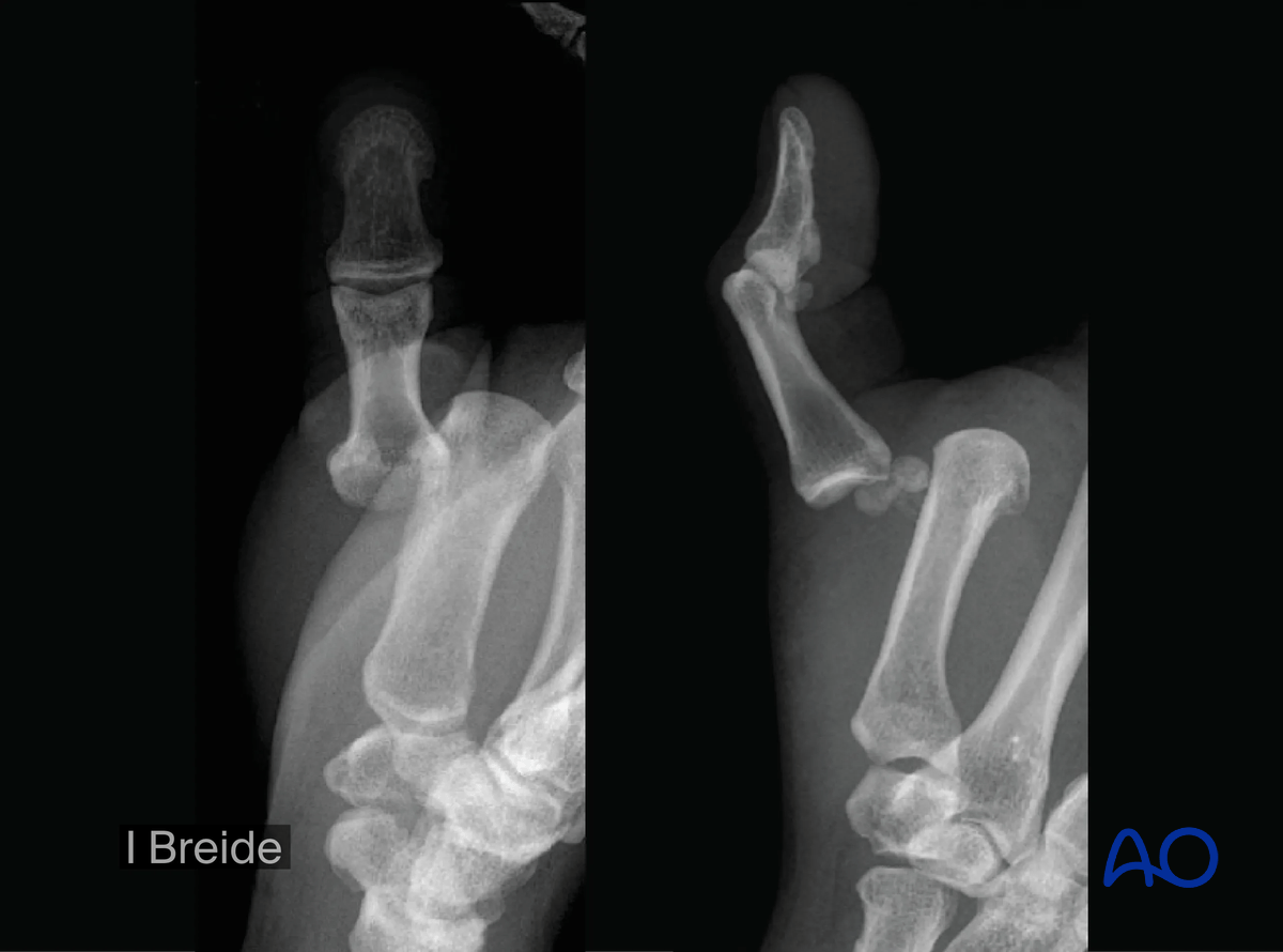
Dislocation of the 1st metacarpophalangeal joint
Description
Metacarpophalangeal (MCP) joint dislocations of the thumb are classified as AO/OTA 70E1.1[5].
Dislocations are most common in the sagittal plane with dorsal dislocations being the most frequent.
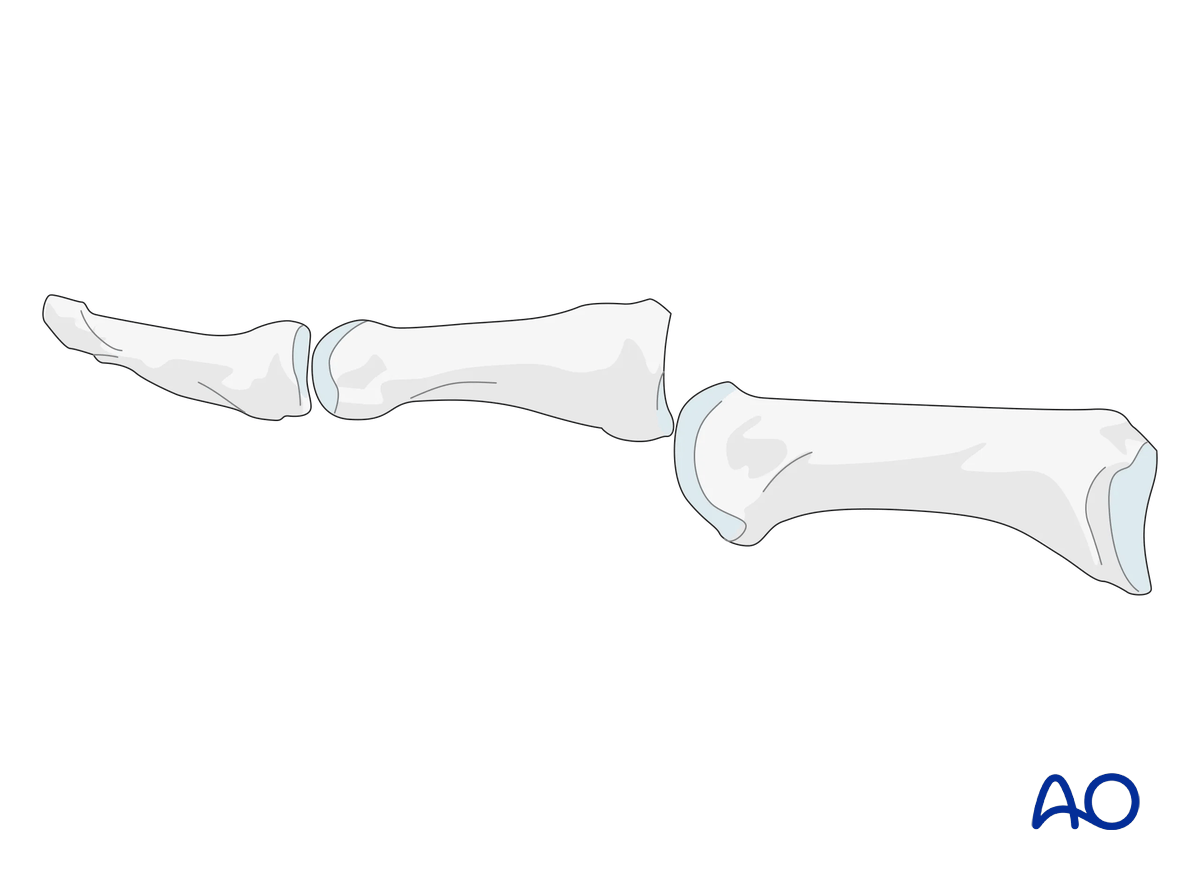
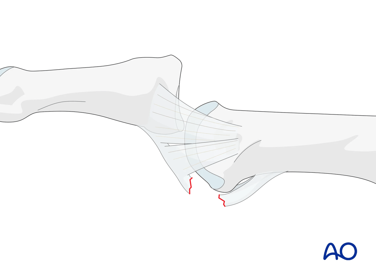
The volar plate is often ruptured in an MCP joint dislocation.
At the MCP joint, the volar plate ruptures proximally due to the lack of the check rein ligaments. At the IP joint, the volar plate ruptures distally.
Therefore, soft-tissue interposition is more likely with an MCP joint dislocation.
Recognizing subluxation
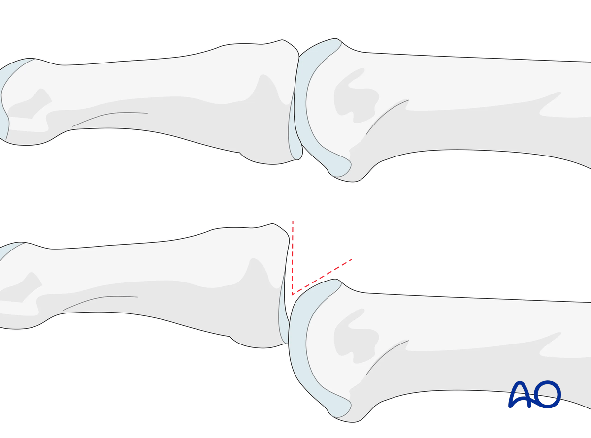
In the lateral view, the proximal phalanx and metacarpal should be collinear. Any axial malalignment is a clear indication of subluxation.
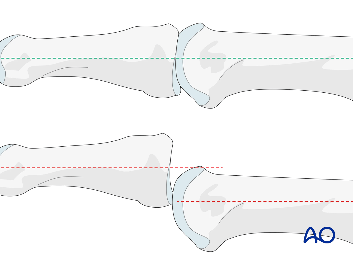
Another indication of subluxation is the presence of a so-called V-sign in the lateral x-ray.
Imaging
Dorsopalmar (PA) and lateral x-rays should be taken.
See the chapter on patient assessment on how to take the x-ray views.