

Patient assessment
1. Introduction
Hand surgery involves a multi-specialty approach to the assessment, treatment, and aftercare of hand trauma. Therefore, the specialized teams should be involved early on according to the specific injury requirements.
A diagnosis is made based on the history, the mechanism of the trauma, clinical examination, and by x-rays in two planes.
2. Clinical assessment
Check for:
- Swelling
- Open wound
- Loss of sensation in the thumb (indicating nerve injuries)
- Perfusion (reperfusion of more than 2 seconds may indicate vascular injuries)
General assessment of hand and wrist
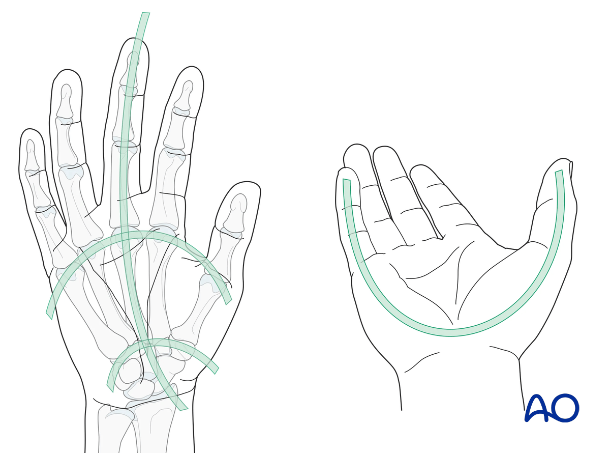
Check for anatomical architecture (position and relation) of the whole hand:
- Longitudinal arc
- Metacarpal arc
- Oblique arcs (with thumb in opposition)
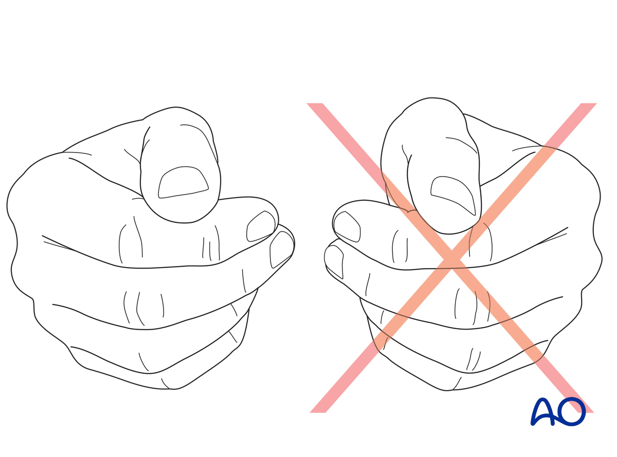
Check for malrotation of the thumb by comparing the nail plates to the contralateral thumb with the hands in resting position.
Assessment of the tendon function
Extensor pollicis longus (EPL)Check retropulsion of the thumb with the hand placed on a table.
Check the strength of MCP joint extension. Weakness may indicate isolated EPB lesion.
Check for deformity of the bone.
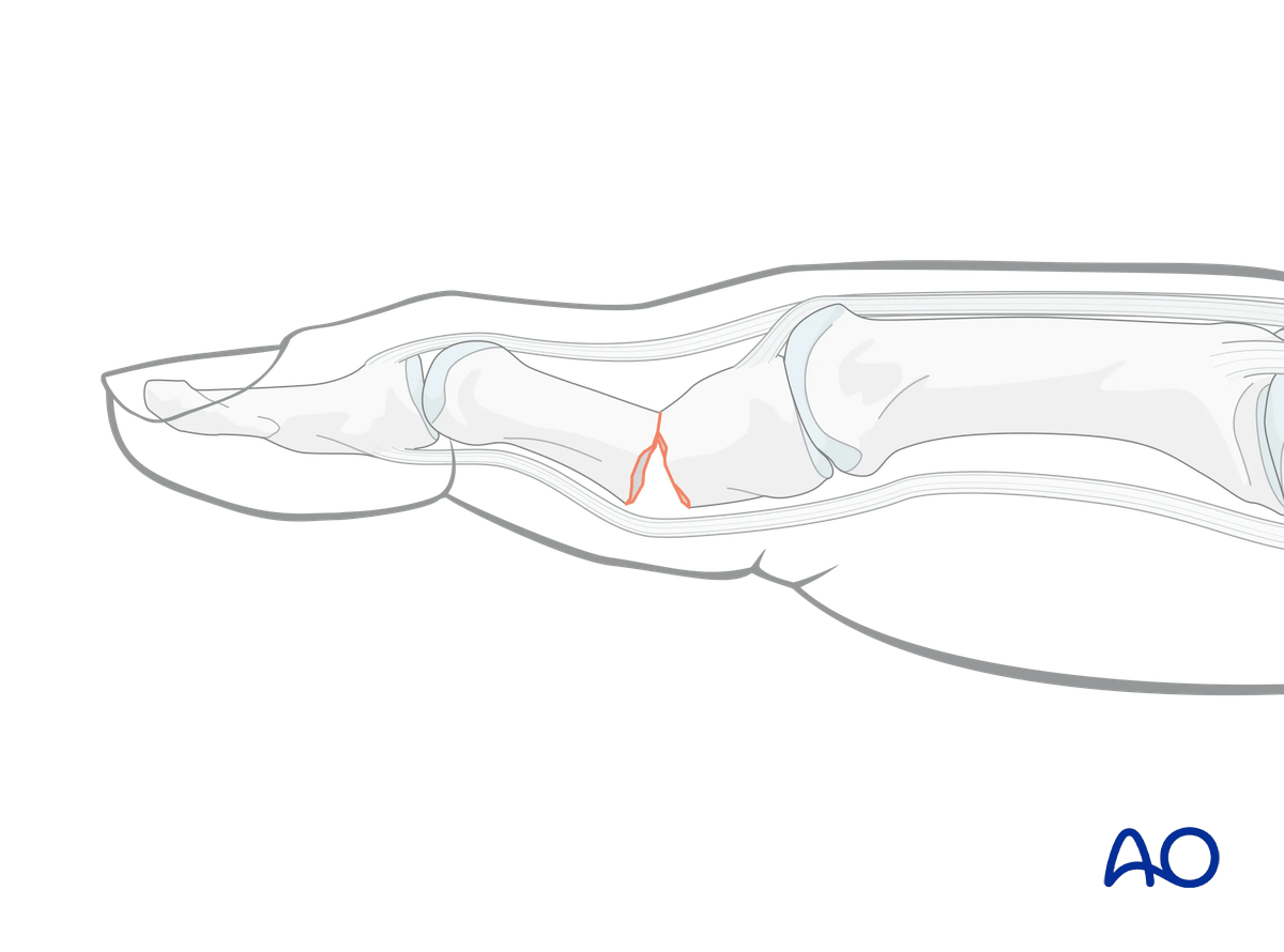
In a metacarpal fracture, the proximal fragment may be pulled dorsally resulting in a flexion deformity.
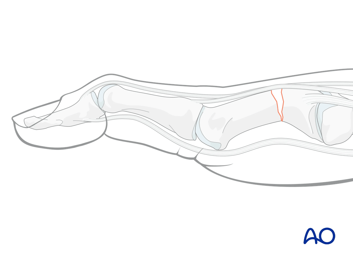
In a proximal phalangeal fracture, the proximal fragment may be pulled palmarly resulting in an extension deformity.
Check the metacarpophalangeal (MCP) and interphalangeal (IP) joint stability. Compare this with the contralateral finger.
Lack of lateral stability indicates injury to the collateral ligaments or their bony attachments.
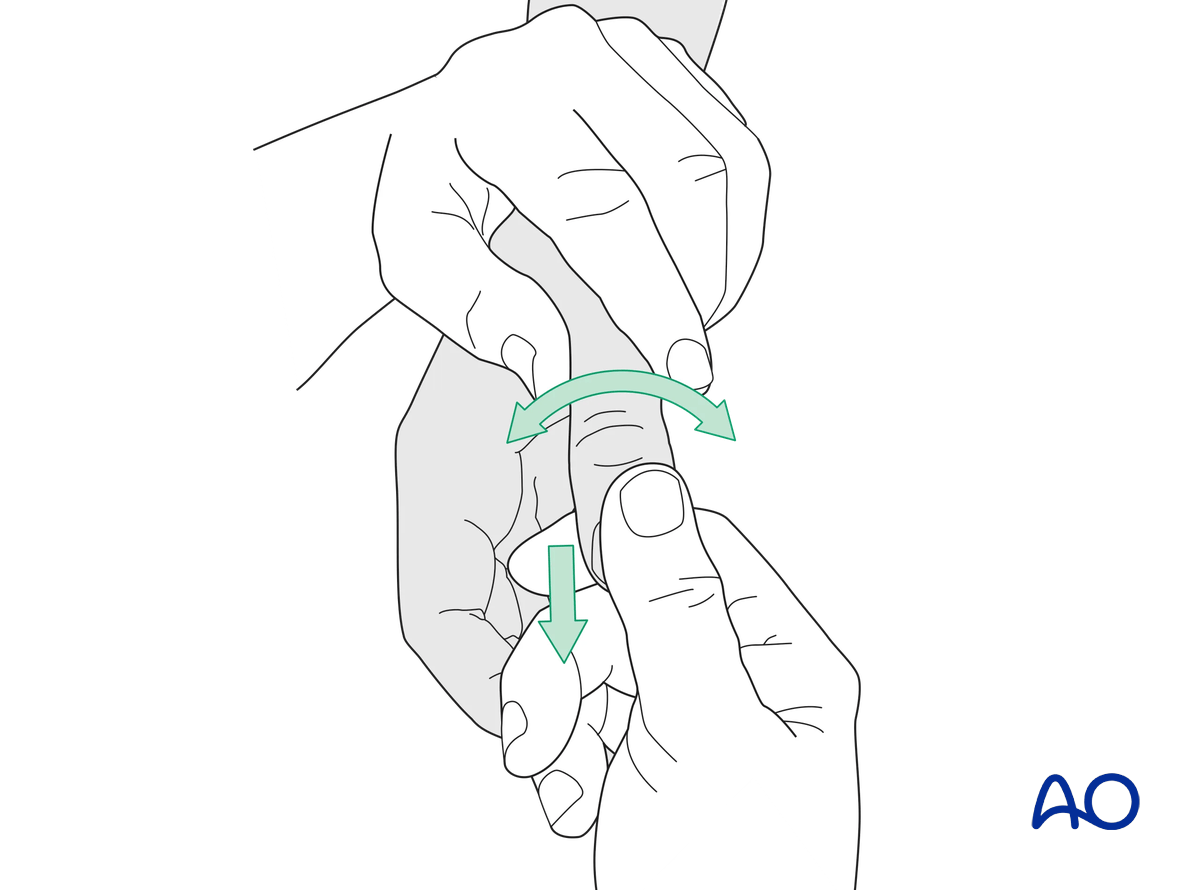
3. Diagnosis of a skier’s thumb
After injection of a local anesthetic into the MCP joint, stress testing is performed in 30 flexion. This is necessary to exclude any influence of the intact volar plate on stability when tested in full extension. Compare the test with the uninjured side. More than 15° difference and a soft end point indicate a complete rupture of the ligament.
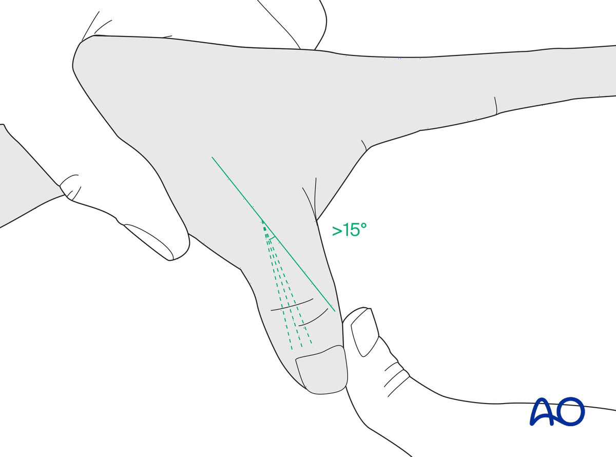
4. Radiological evaluation
X-rays
Dorsopalmar (PA) view of the whole hand and lateral view of the thumb are needed for diagnosis. In base fractures, an oblique view may be helpful.
All views need to be inspected to get information about the fracture pattern, eg, the orientation of an oblique fracture plane.
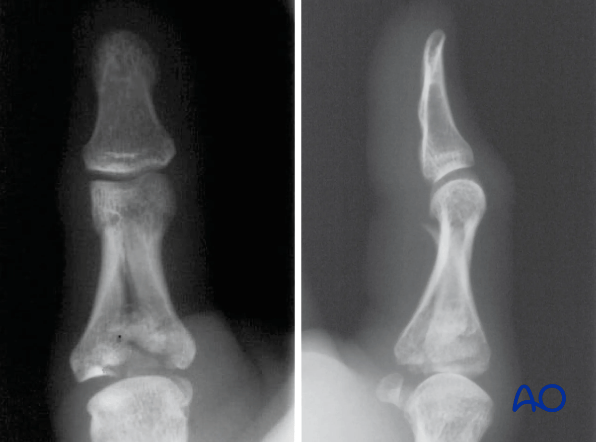
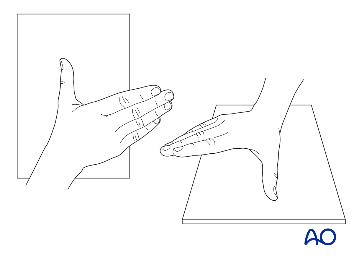
To obtain the true PA view (Robert’s view), rotate the hand internally with the thumbnail resting against the cassette.
The x-ray should show open IP, MCP, and CMC joints.
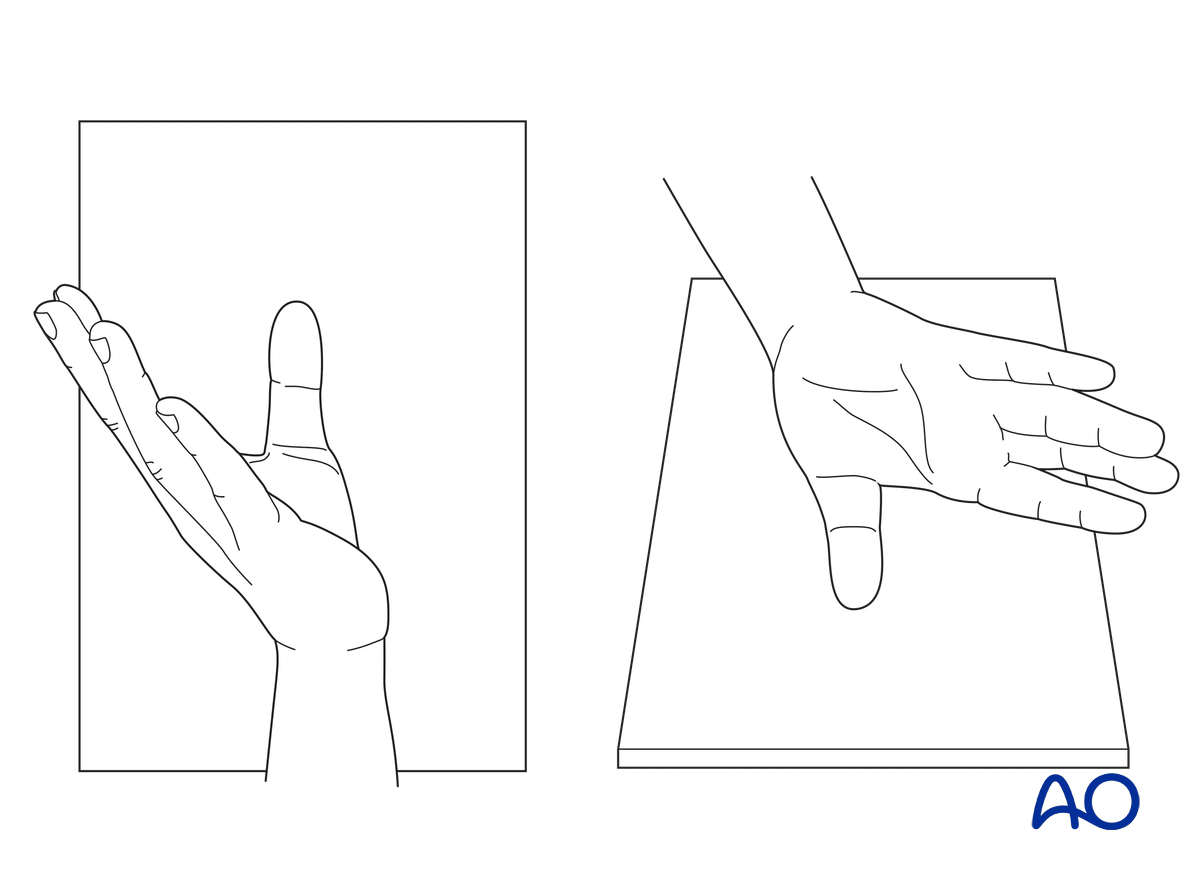
To obtain the true lateral view (Bett’s view), flex the hand and slightly rotate it externally.
The x-ray should include the CMC, MCP, and IP joints.
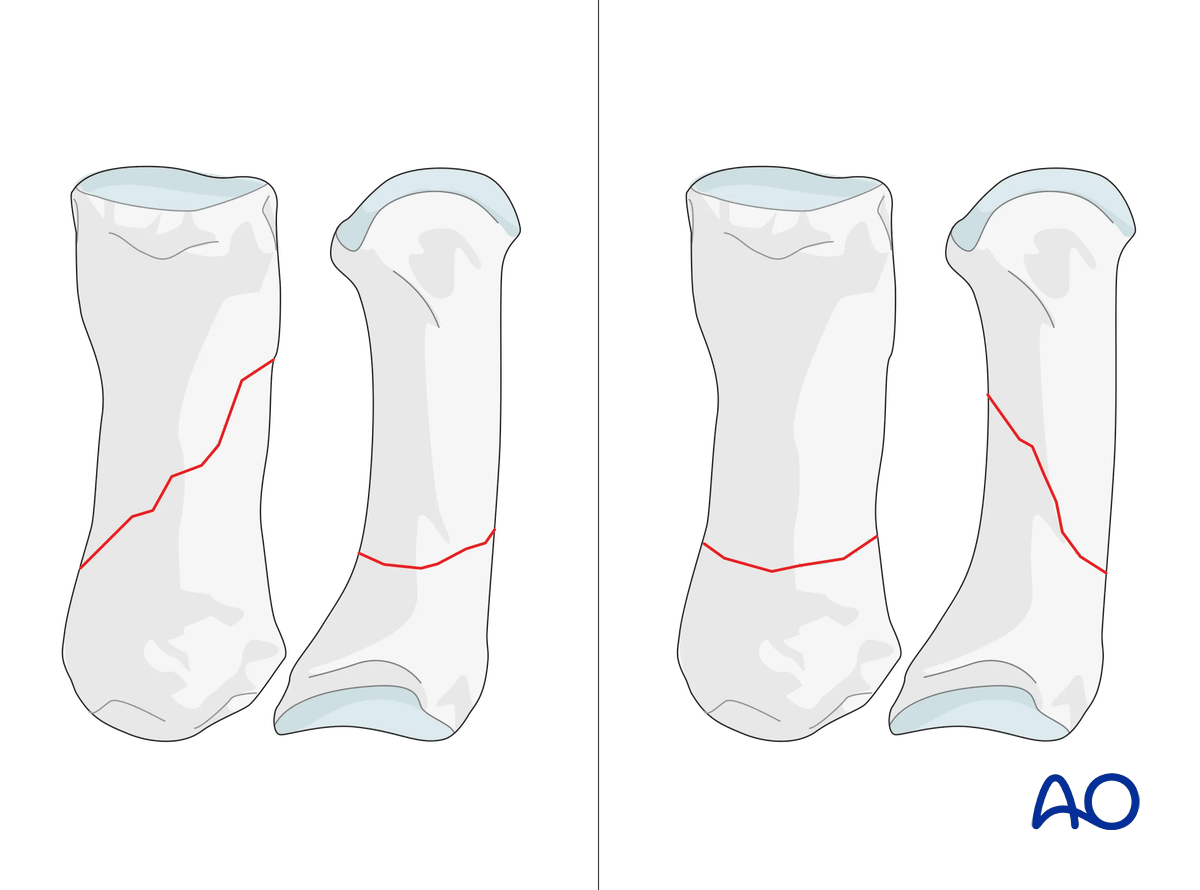
Obliquity of the fracture is possible either in the plane visible in the AP view or the plane visible in the lateral view. Always confirm the fracture configuration with views in both planes.
Ultrasound
Ultrasound evaluation may help define suspected ligament and tendon injuries.
CT imaging
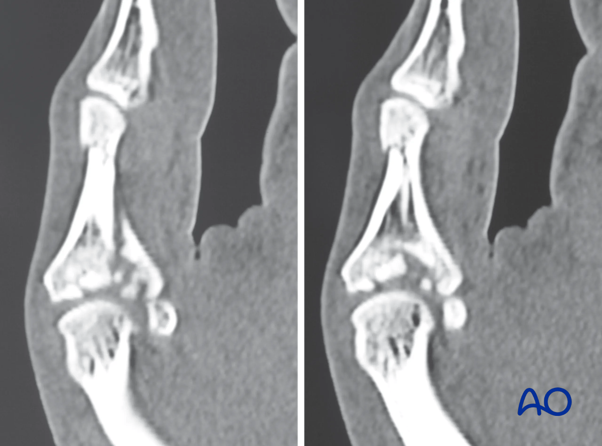
CT imaging is required for impacted intraarticular fractures to evaluate the fragmentation and plan for reconstruction.
MRI
Isolated ligamentous injuries may be seen in an MRI.


