

Nonoperative treatment
1. General considerations
This fracture is generally unstable and often requires open reduction and stable fixation.
Stable undisplaced fractures or in frail patients, conservative treatment may be considered.
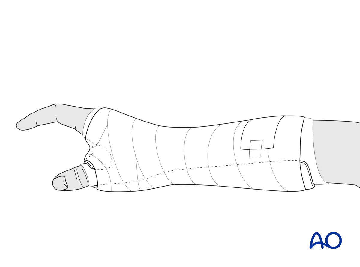
MCP and wrist joints should be immobilized with a thumb spica or thermoplastic splint.
Any axial displacement should be reduced to gain full length of the metacarpal.
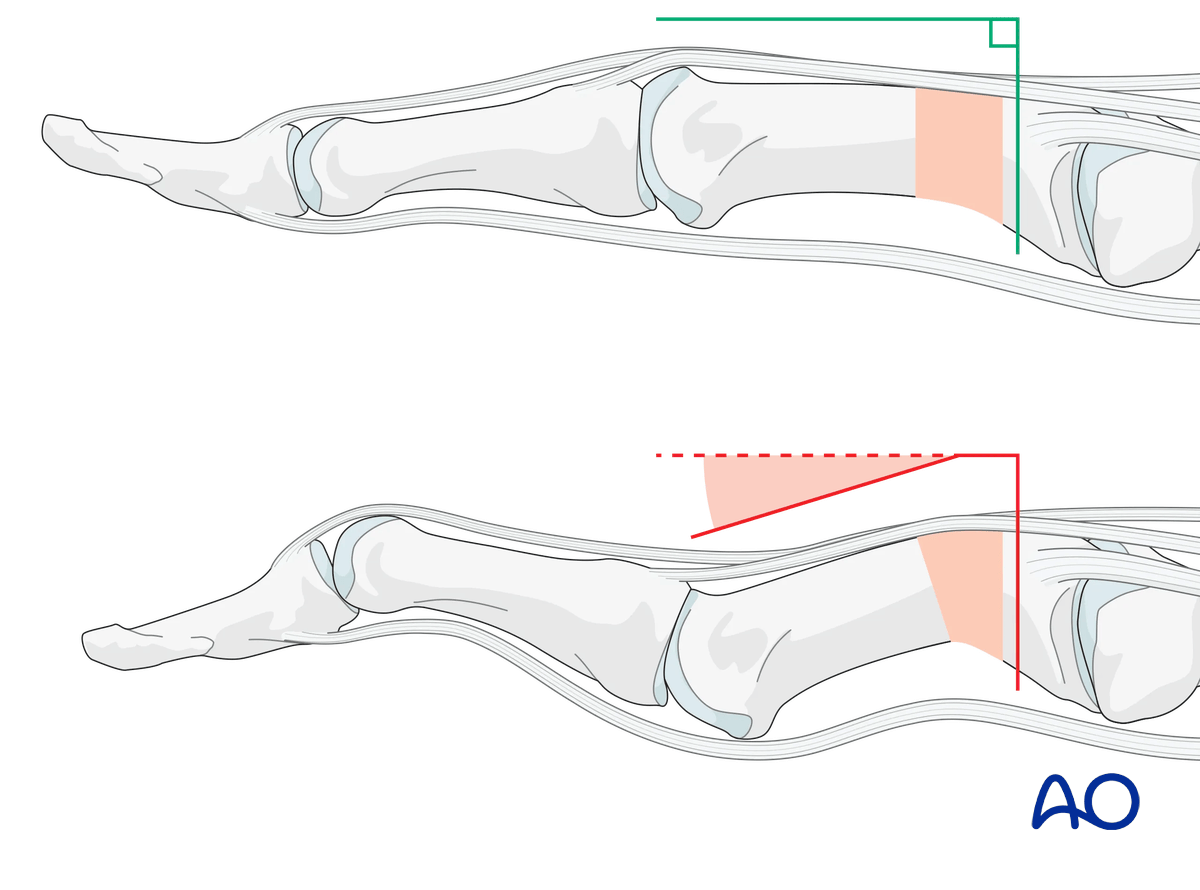
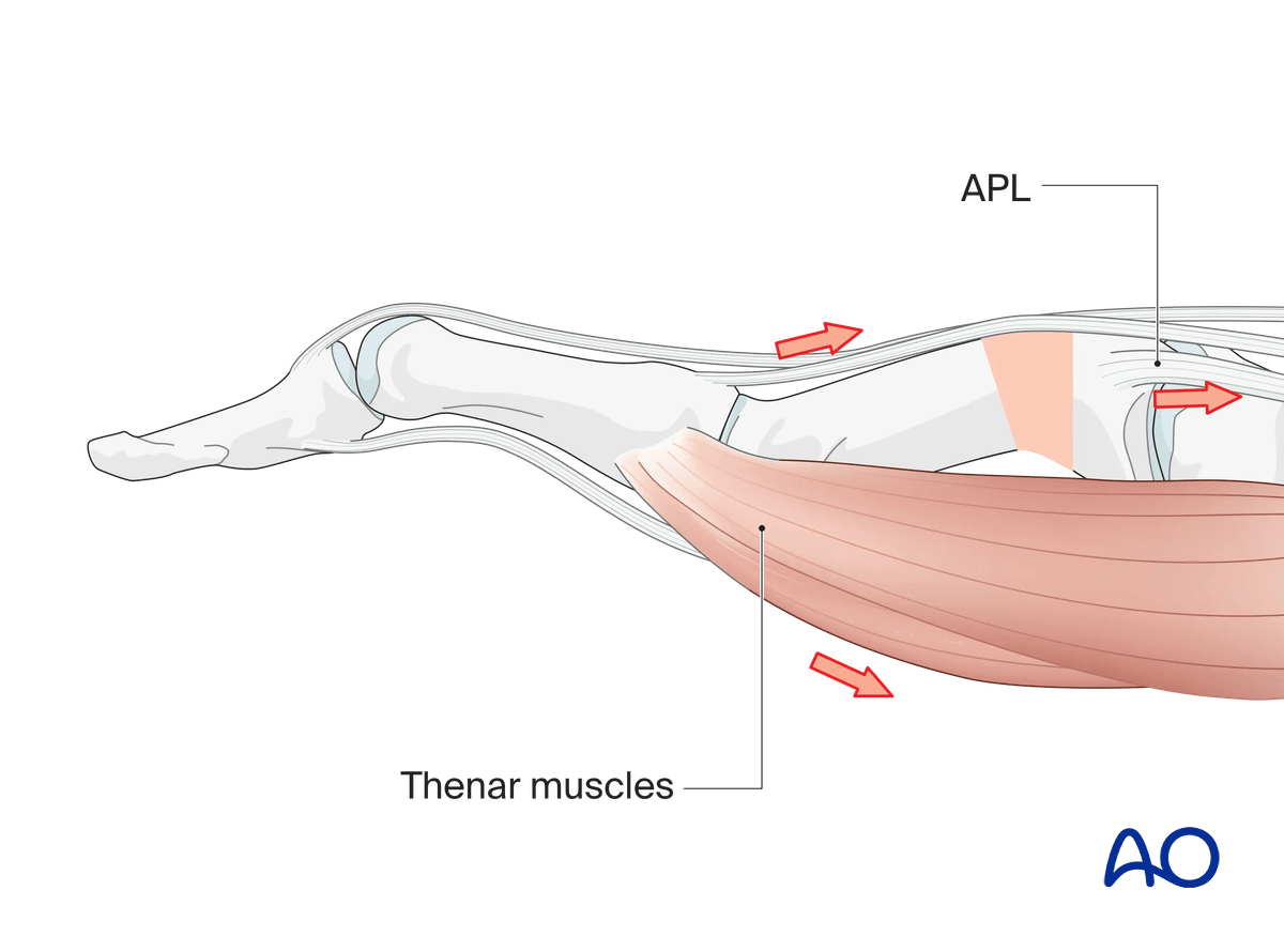
This illustration shows the effect of the combined dynamic action of APL and the thenar muscles across malalignment.
The relative shortening of the metacarpal allows the extensor lag due to relative lengthening of the EPL.
The APL pulls the proximal fragment into extension.
The thenar muscles pull the distal fragment into flexion.
This results in flexion of the IP joint and hyperextension of the MCP joint (Z-deformity).
2. Patient preparation
Anesthesia
To reduce pain during closed reduction and cast application, local anesthesia (nerve block of the median and superficial radial nerve or WALANT) may be administered.
See also preoperative preparation.
3. Reduction
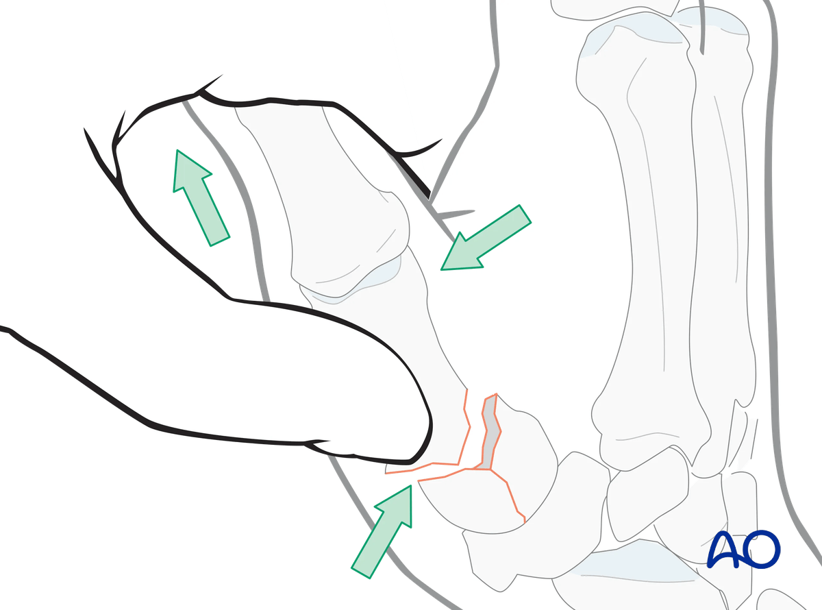
As there is usually a flexion deformity, reduction of the diaphyseal fragment to the articular fragments can be performed with axial traction on the thumb and simultaneous pressure over the dorsal aspect of the basal diaphysis near the fracture.
4. Splint application
Immobilize the hand and wrist in a well-padded thumb spica splint with the wrist slightly extended and the 1st MCP joint immobilized in a position of abduction.
Metacarpal and Phalanx—Fractures and Soft-tissue Injuries—Thumb Spica
Paul Demmer, Doron JM Frantzen, Nikolaas GJ Maritz
The thumb spica is a circular plaster cast used to immobilize the thumb. In this presentation the application of the thumb spica will be demonstrated.
Maintaining reduction
During the application of the splint, it is important to maintain thumb abduction by pressure over the palmar aspect of the first metacarpal head and the dorsal aspect of the 1st metacarpal base.
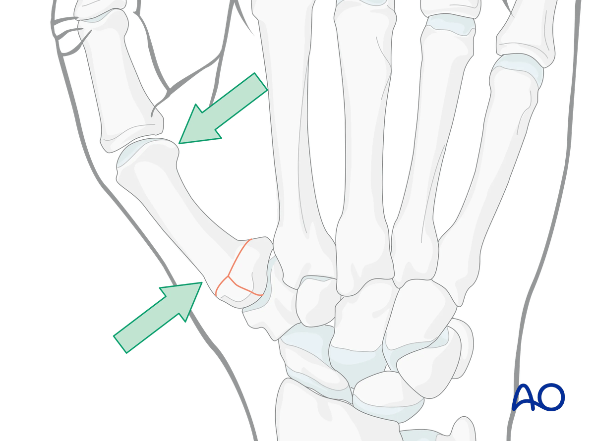
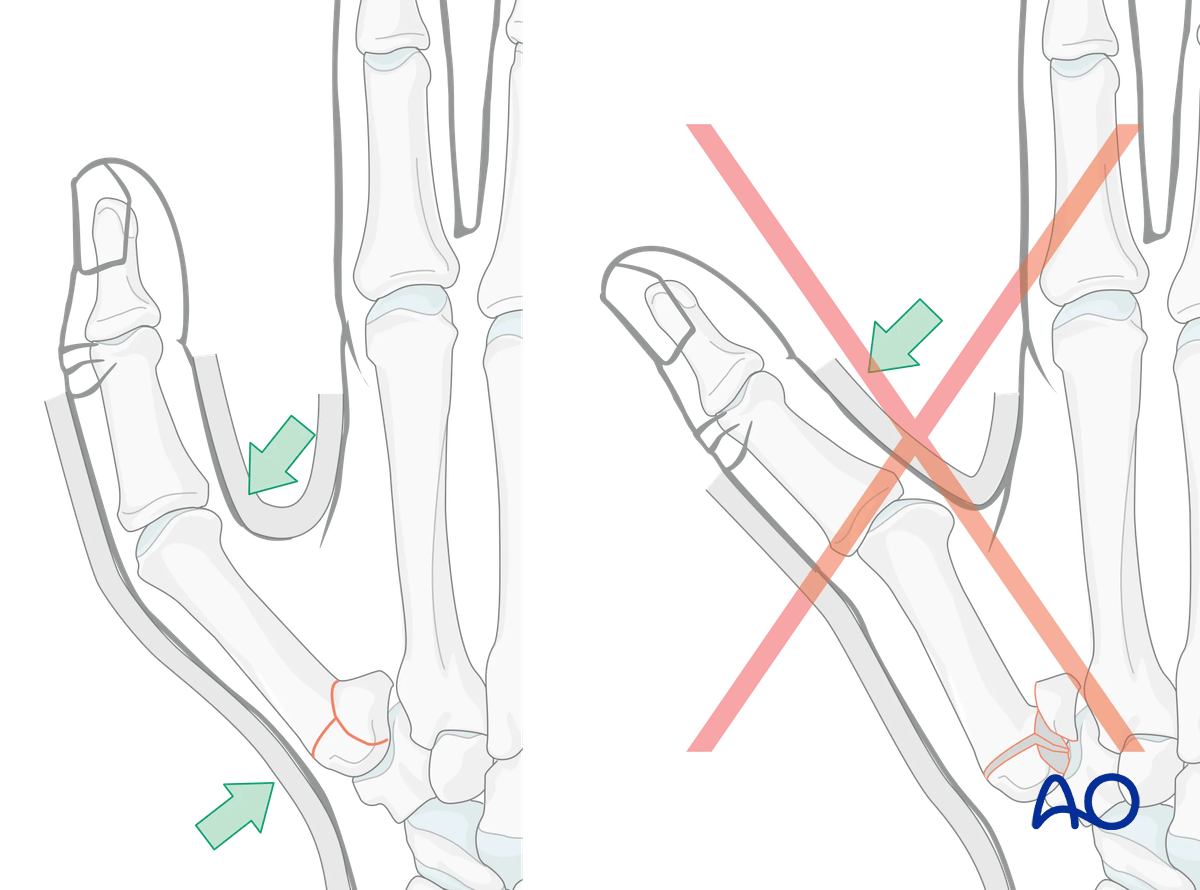
Splinting
Protect the skin of the arm and hand with padding/cotton sleeve to avoid pressure sores, especially on the 1st webspace, distal ulna, and styloid process of the radius.
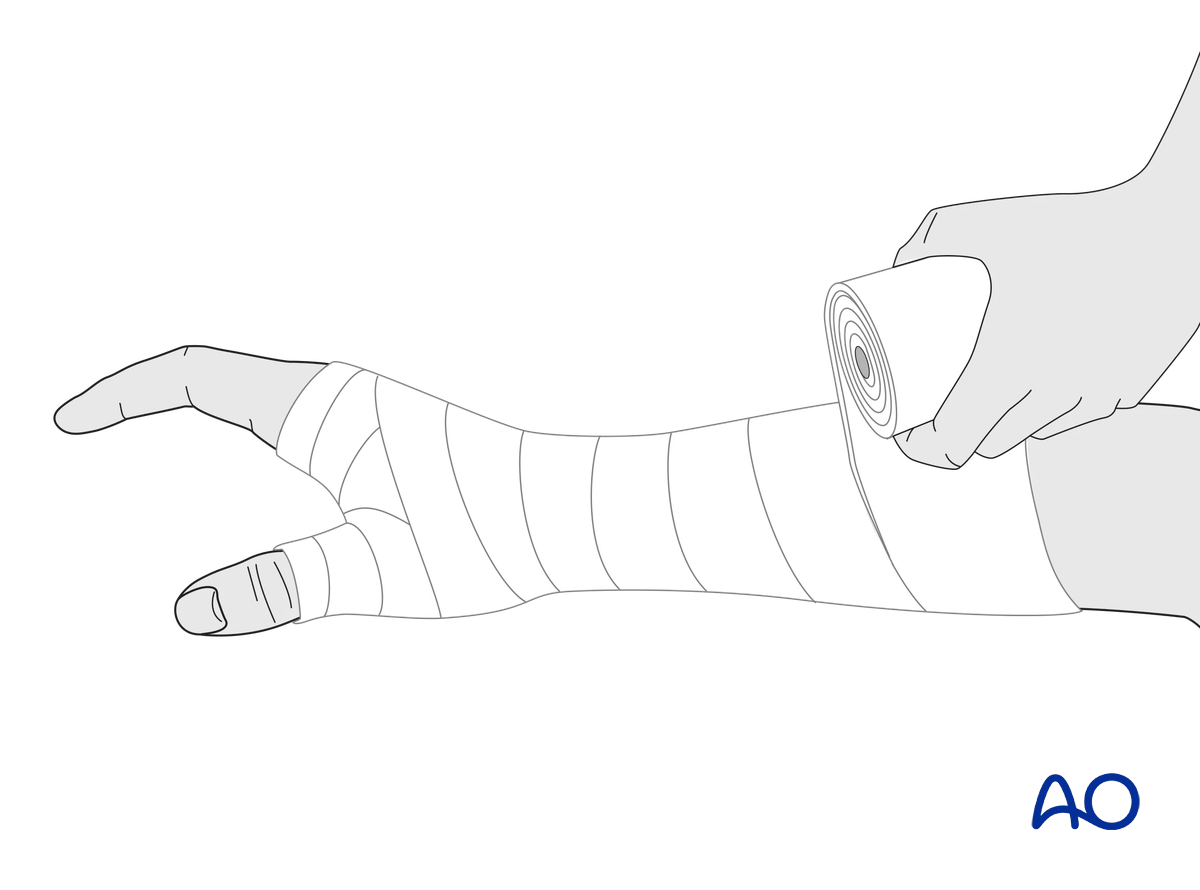
Create a splint of several layers of cast bandage depending on the specific type of bandage available. It should cover the palmar half of the forearm, wrist, and thumb.
The wrist should be in neutral or slight extension.
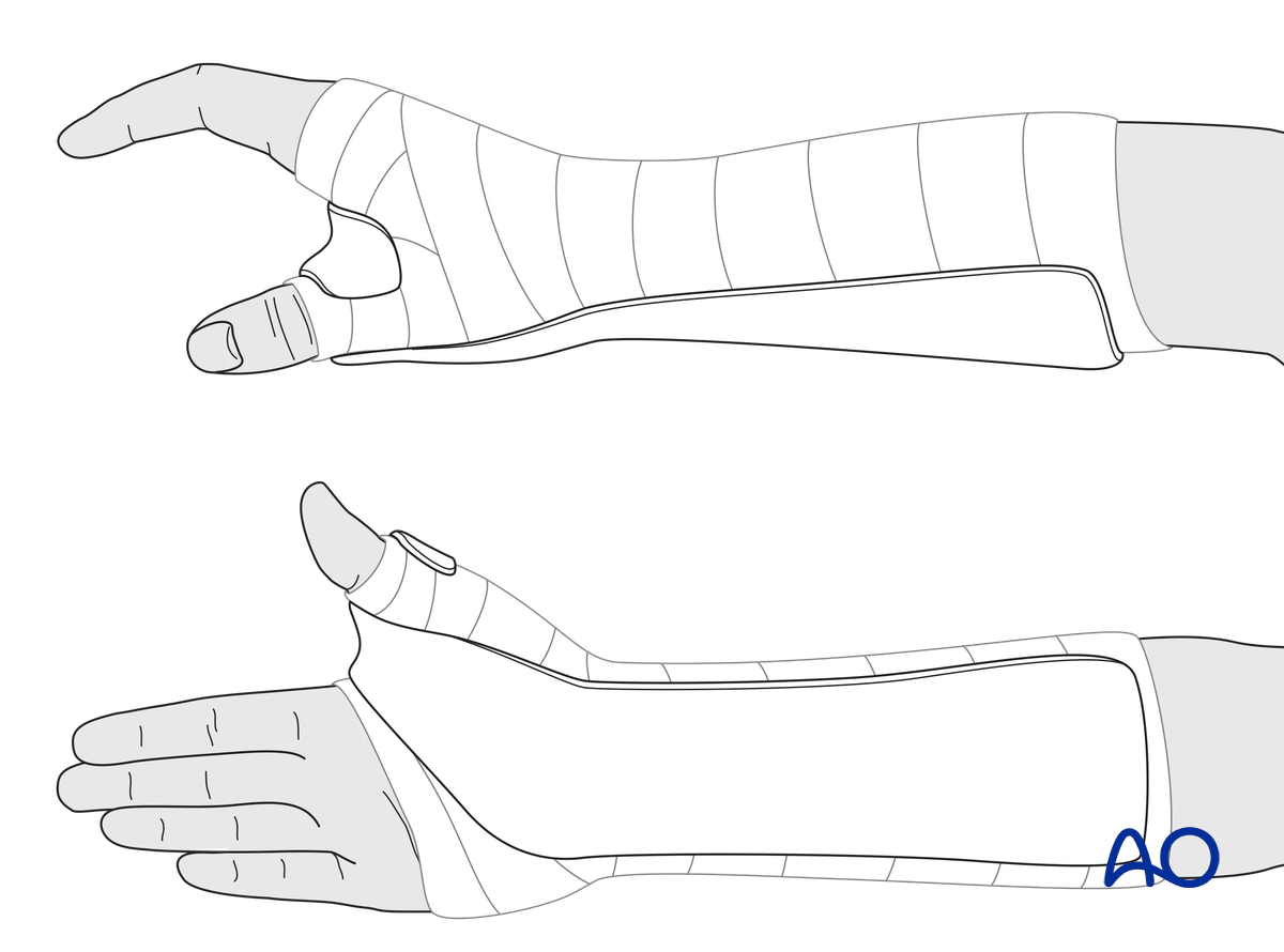
Make sure that the splint does not extend too far distally, either at the level of the finger metacarpophalangeal (MCP) joints, or the interphalangeal joint of the thumb. The splint must not limit complete flexion of these joints.
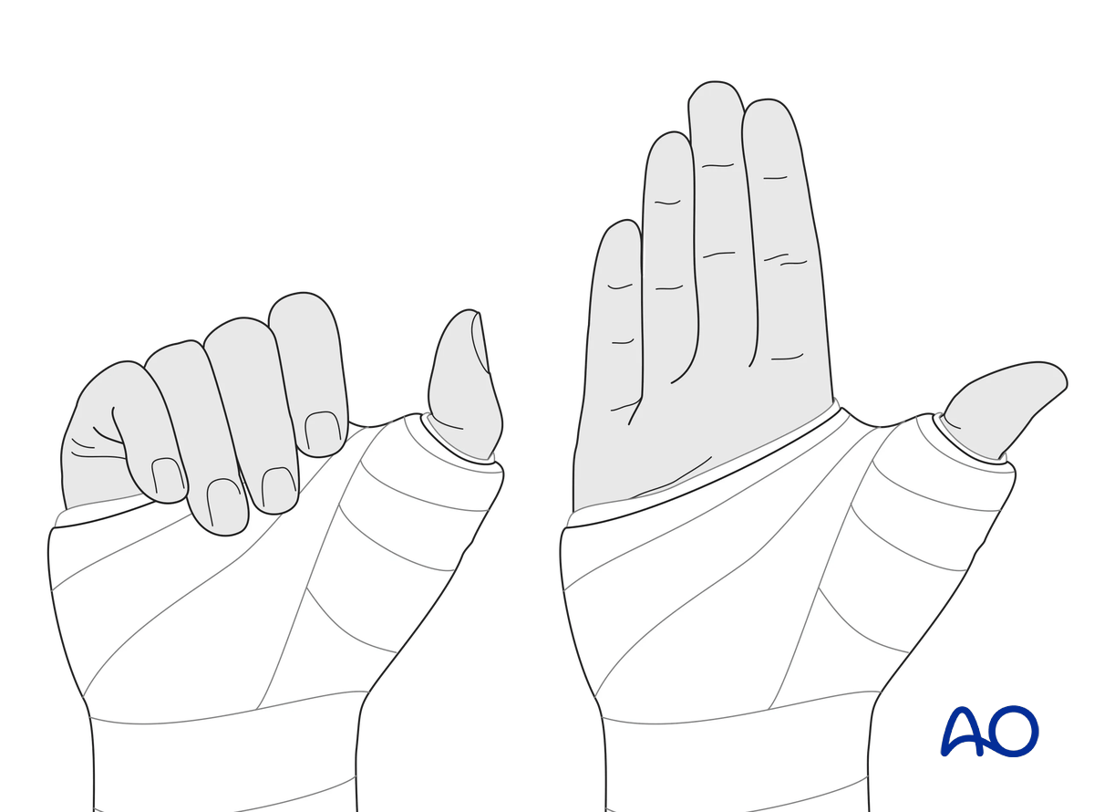
The splint is held in place with an elastic bandage. The bandage should not be overtightened, especially at the level of the wrist joint and 1st webspace, to avoid pain and excessive swelling of the hand.
Direct skin contact with adjacent fingers should be prevented by placing gauze pads between them.
The patient should be instructed to add another elastic bandage if necessary.
5. Aftercare
Healing phases
The aftercare can be divided into four phases of healing:
- Inflammatory phase (week 1–3)
- Early repair phase (week 4–6)
- Late repair and early tissue remodeling phase (week 7–12)
- Remodeling and reintegration phase (week 13 onwards)
Full details on each healing phase can be found in the chapter Postoperative treatment of thumb injuries.
Management of swelling
The arm should be actively elevated to help reduce any swelling.
Mobilization
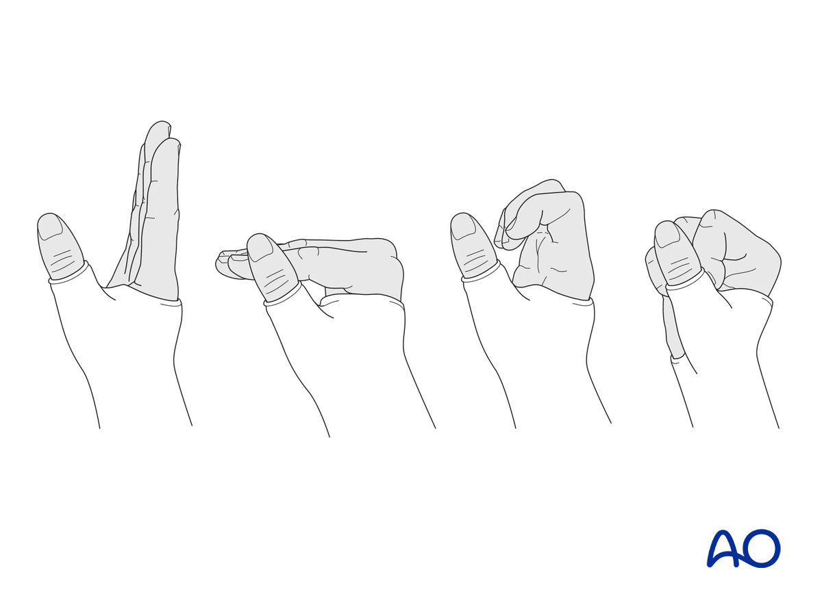
To prevent joint stiffness, the patient should be instructed to begin active motion (flexion and extension) of all nonimmobilized joints of the hand immediately.
Follow-up
X-ray checks of fracture position should be performed immediately after the splint has been applied.
Follow-up x-rays with the splint should be taken after 1 week and after 2 weeks if there is doubt about maintenance of reduction.
Splinting is continued until about 5–6 weeks after the injury. At that time, an x-ray without the splint is taken to confirm healing, and manual pressure on the fracture site should be pain-free.
Return to daily activities
Heavy manual demand and all activities involving a strong grip are not permitted until complete fracture healing (usually after 3 months).


