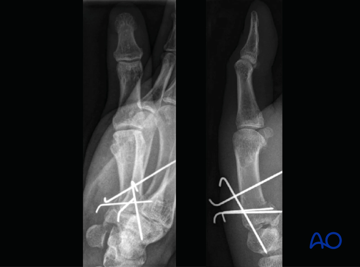
K-wire fixation
1. General considerations
If closed reduction is successful, indirect stabilization with K-wires may be performed to counteract the action of abductor pollicis longus (APL) combined with intermetacarpal transfixation for rotational stabilization.
If the fracture configuration allows, direct K-wire fixation may be performed.
It is important to keep the first webspace open.
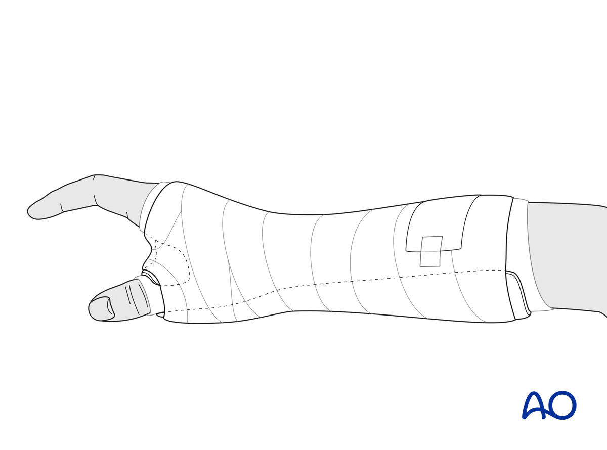
MCP and wrist joints should be immobilized with a thumb spica cast or thermoplastic splint until K-wire removal.
2. Reduction
Any axial displacement should be reduced to gain full length of the metacarpal.
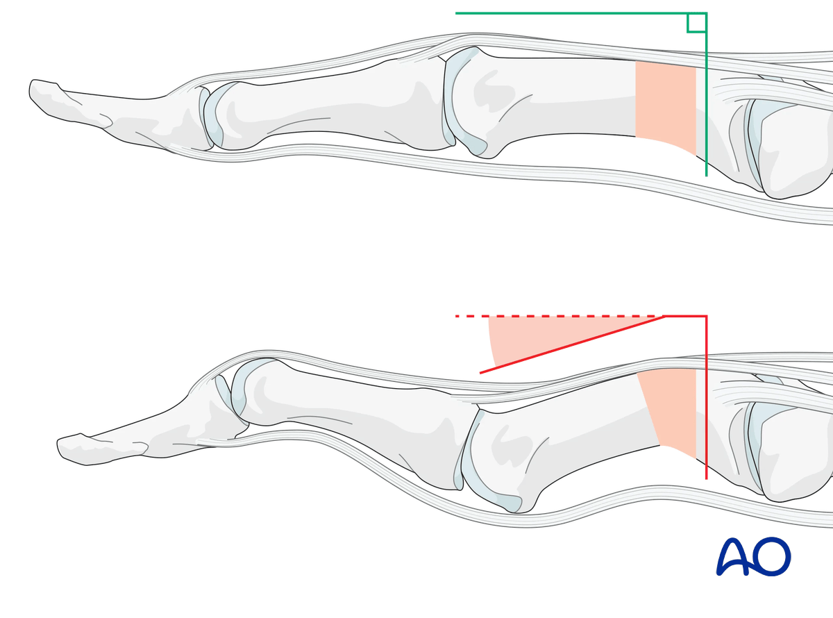
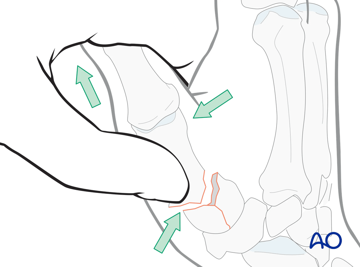
As there is usually a flexion deformity, reduction of the diaphyseal fragment to the articular fragments can be performed with axial traction on the thumb and simultaneous pressure over the dorsal aspect of the basal diaphysis near the fracture.
3. Protecting soft tissues by blunt dissection
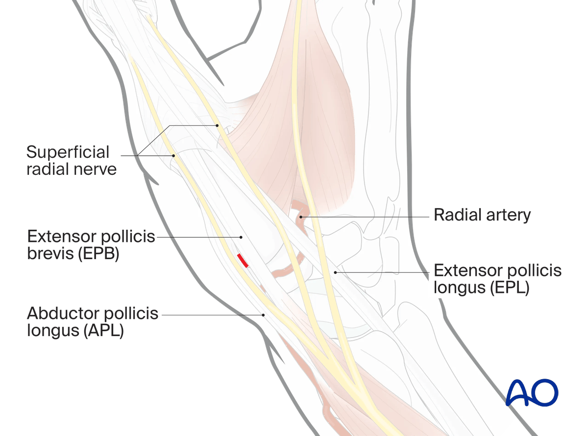
Perform a dorsal stab skin incision over the base of the thumb. Use gentle blunt dissection with artery forceps to develop a tract down to the periosteum before insertion of the K-wire.
Identify and protect the cutaneous branches of the superficial radial nerve and the extensor tendons.
- Damage to the cutaneous nerve(s) can cause a painful neuroma.
- Damage to the tendons can cause adjacent adhesions which limit tendon excursion.
X-rays to check that the articular surface is anatomically reduced.
If reduction is not anatomical, proceed to an open operation.
4. K-wire stabilization
Use 1.2–1.6 mm K-wires.
They may be cut off below the surface of the skin, or left protruding through the skin, in which case the wires are bent over.
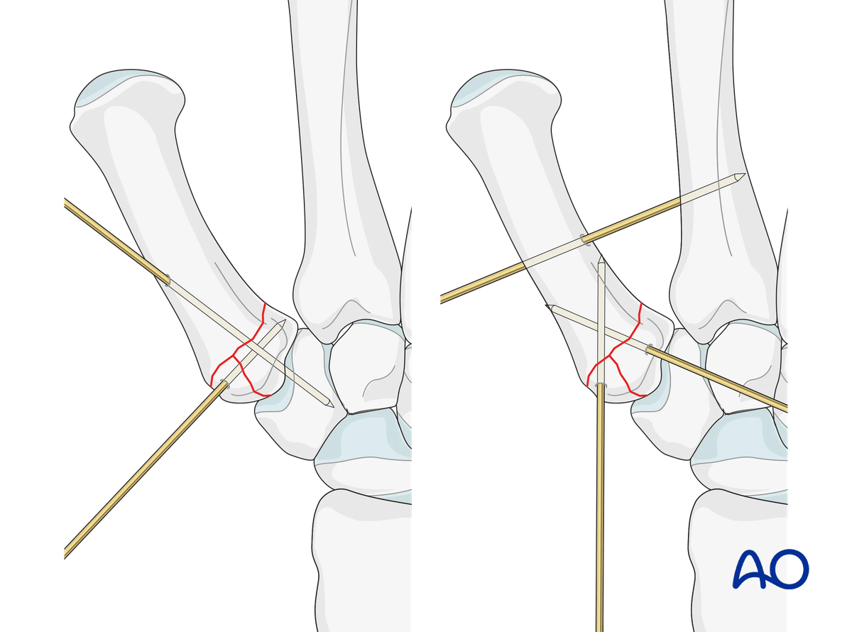
If the fracture configuration and size of the fragment allow, insert K-wires through the articular fragment into the diaphysis.
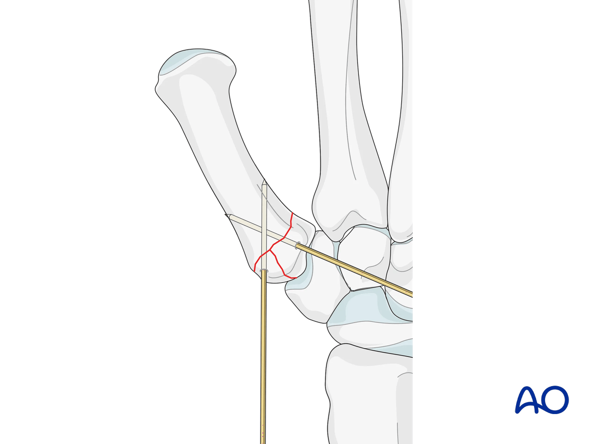
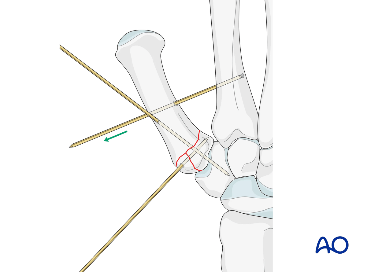
There are several possible K-wire constructs to counteract the pull of the APL and ensure fracture stabilization:
- Transfixation of the CMC joint
- Intermetacarpal transfixation
- Combination of CMC joint and intermetacarpal transfixation (distal to the Bennett fragment)
5. Final assessment
Confirm correct rotational alignment by clinical examination.
Image intensification may be used to confirm anatomical reduction and correct placement of implants in two views.
6. Splinting
4–6 weeks of immobilization in a thumb spica splint are necessary.
7. Aftercare
Healing phases
The aftercare can be divided into four phases of healing:
- Inflammatory phase (week 1–3)
- Early repair phase (week 4–6)
- Late repair and early tissue remodeling phase (week 7–12)
- Remodeling and reintegration phase (week 13 onwards)
Full details on each healing phase can be found in the chapter Postoperative treatment of thumb injuries.
Management of swelling
The arm should be actively elevated to help reduce any swelling.
Cleaning protruding K-wires
In the case of protruding K-wires, instruct the patient how to clean the wound every second day. For this, a window must be cut in the cast, or splint, at the sites of the K-wires.
Functional exercises
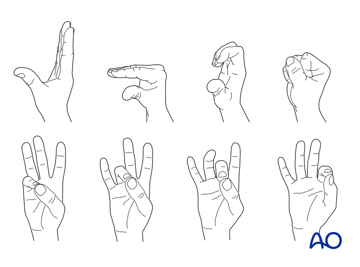
During the immobilization, flexion and extension exercises of the MCP and interphalangeal joints of the nonoperated fingers are performed. Only the IP joint is mobilized in the thumb.
Opposition of thumb to the other fingers is performed after K-wire removal.
Weight bearing should be avoided until fracture healing is confirmed.
Follow-up
See the patient after 2 weeks of surgery for suture removal.
After 6 weeks, an x-ray should be taken to confirm healing.
Implant removal
After 4–6 weeks, the K-wires are removed as an outpatient procedure.
8. Case
X-rays of a complete articular fracture of the proximal end segment of the thumb metacarpal (Rolando fracture)
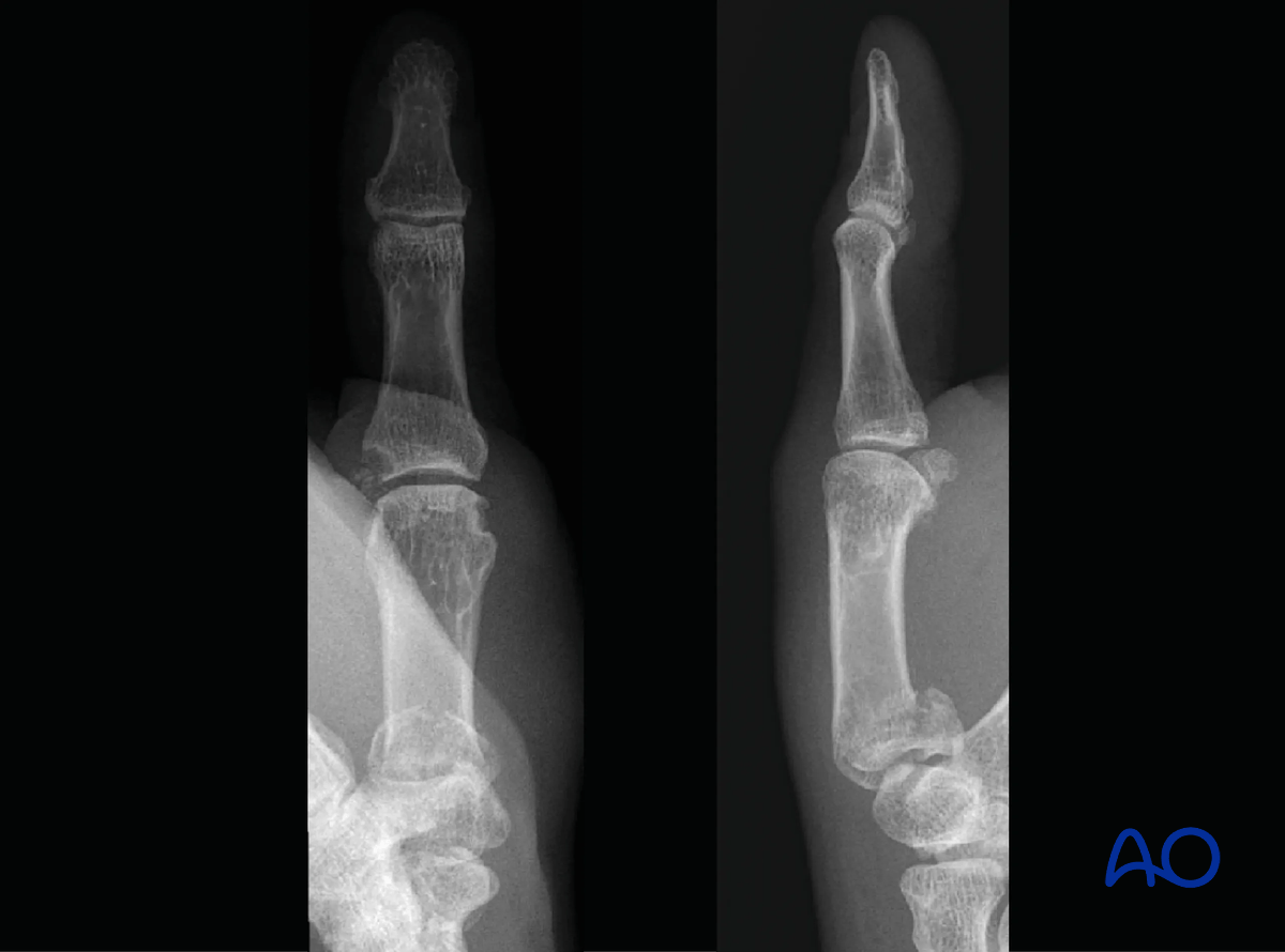
X-rays of the final construct with K-wire fixation