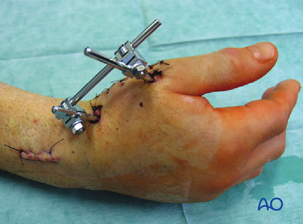
External fixation
1. Principles
Introduction
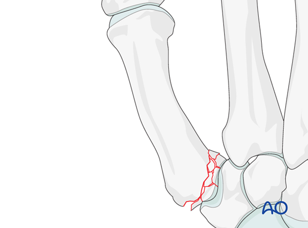
The term “Rolando’s fracture” is often used to describe multifragmentary intraarticular fractures of the thumb base.
If the fragments are numerous and small it is not advisable to attempt internal fixation, because of the risk of fragment devascularization and the technical difficulty of fixing small fragments.
In such cases, external fixation with gentle distraction across the joint maintains reduction of the articular surface and preserves the length of the first metacarpal.
Bone graft of any metaphyseal defect is obligatory.
Diagnostic radiology
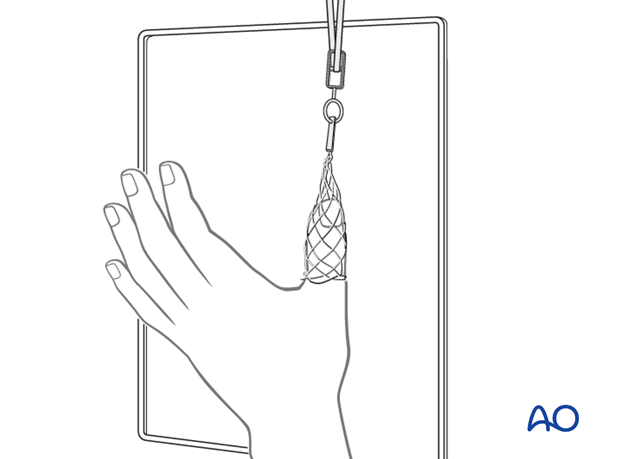
Distract the thumb for standard x-rays, or use CT scans for defining the fracture pattern.
Choice of approach
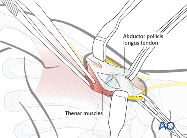
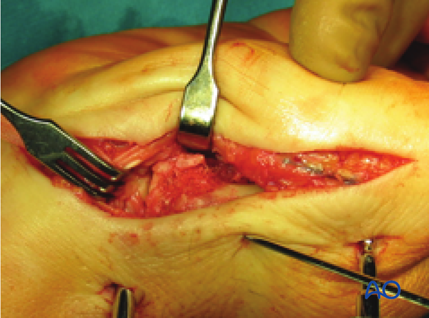
Open reduction is done through the radiopalmar approach. This is combined with a capsulotomy of the first carpo-metacarpal joint, which helps in the restoration of the articular surface.
Teaching video
AO teaching video:The mini external fixator
2. Restoration of the articular surface
Distraction
The use of a small distractor is advisable.
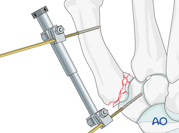
As an alternative, use a small external fixator with a distraction clamp for distraction.
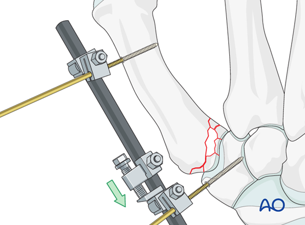
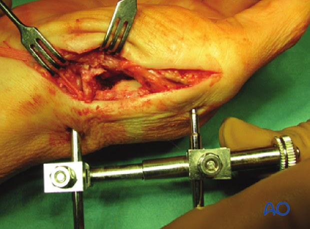
Insert the distal pin into the metacarpal diaphysis.
Then insert the second pin into the trapezium using blunt dissection to protect the soft tissues.
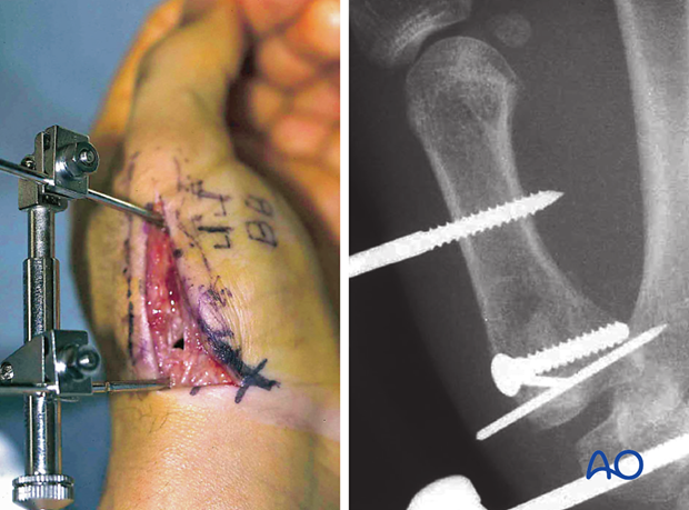
Disimpaction
Distract the carpo-metacarpal joint.
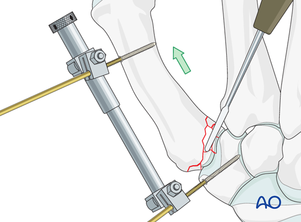
Disimpact intraarticular fragments, using a periosteal elevator and the articular surface of the trapezium as a template.
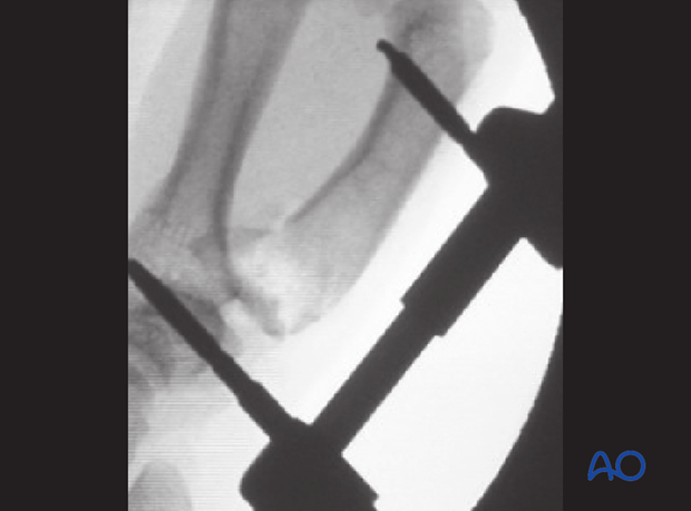
Check the position of the articular surface and the diaphysis using image intensification.
3. Bone graft
Harvest site
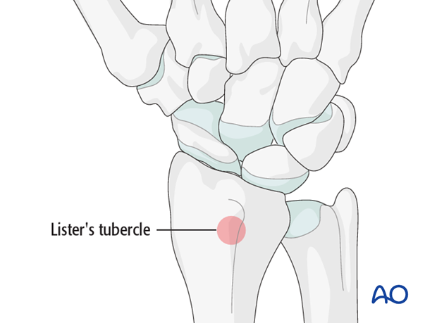
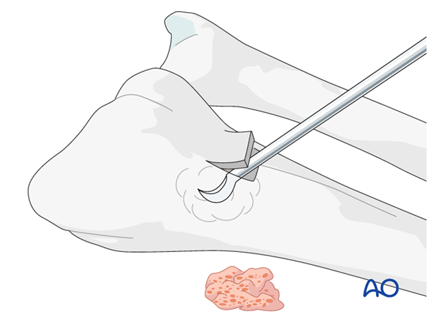
Harvest the bone graft material from the distal radius. A good and safe place for this is just proximal to Lister’s tubercle.
Harvesting
Make a 2 cm long incision proximal to Lister’s tubercle. Retract the tendons of the second compartment radially, and the EPL in an ulnar direction.
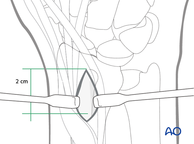
Use a chisel to cut three sides of a square. Hinge up the dorsal cortical flap. After harvesting cancellous bone, replace the “lid” and suture the periosteum and the skin incision.
Insert bone graft
Insert bone graft to fill any subchondral metaphyseal defect.
4. External fixation
Change to external fixator
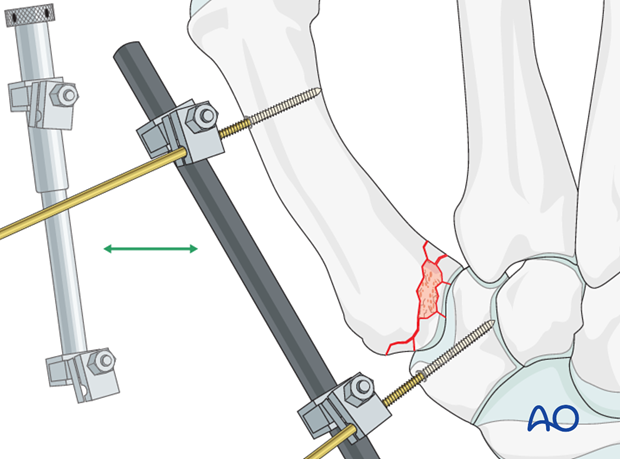
Take an external fixator rod and two clamps and attach them to the pins, between the hand and the distractor body.
With the external fixator maintaining distraction, the distractor body can now be removed.
Option: additional K-wire
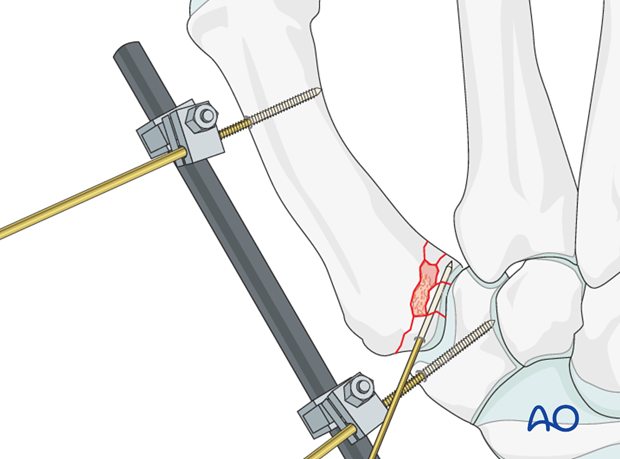
A K-wire can be used to fix larger fragments to each other, or to transfix the joint in order to improve stability.
5. Aftertreatment
The external fixator allows functional aftercare.
Instruct the patient how to clean the pin sites at 2-day intervals.
The external fixator is removed after 4-6 weeks.
After removal, motion of the thumb base is initiated.
Heavy manual demand and all activities involving a strong grip are not permitted until complete fracture healing (usually after 3 months).
