Hinged external fixation following ORIF
1. General considerations
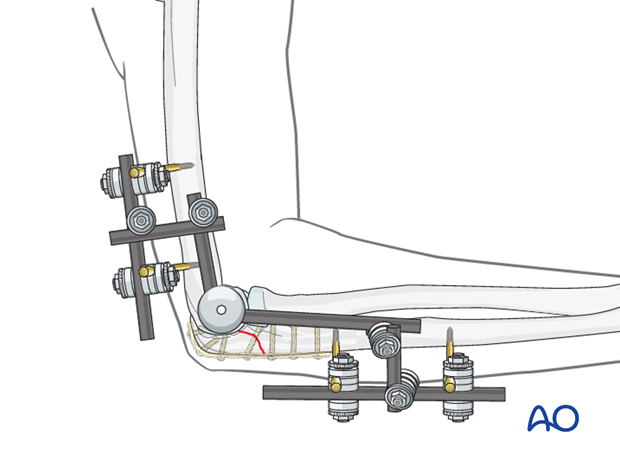
Hinged external fixation can be an important supplement to ORIF and ligament repair for selected complex unstable elbow injuries.
When surgical repair does not provide adequate elbow stability, redislocation or subluxation may occur. External fixation with a hinged device will help keep the elbow reduced while allowing passive and active controlled motion.
The elbow joint closely approximates a simple hinge. Its axis of rotation lies at the centers of the trochlea and capitellum. The fixators hinges are placed along the axis of the joint.
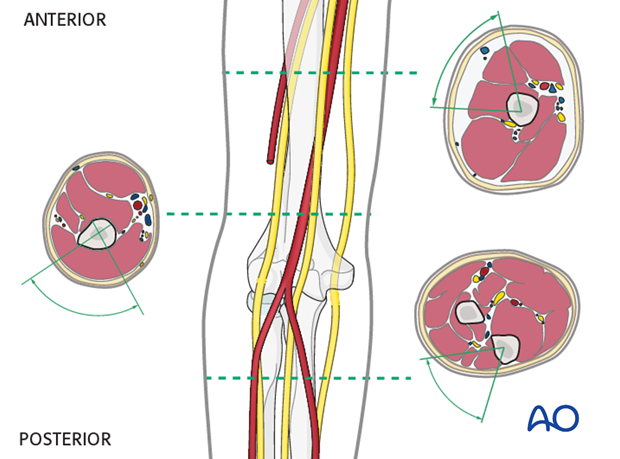
The biggest risk of external fixation of the proximal forearm is iatrogenic neurovascular nerve injury.
Knowing the safe zones and blunt dissection to bone is critical.
2. Positioning
The patient is placed in a supine position:
3. Pin site preparation
Pin placement
For safe pin placement make use of the safe zones and be familiar with the anatomy of the humerus and the forearm.

Soft tissue dissection
Blunt dissection of the soft tissues and the use of small Langenbeck retractors will prevent damage to muscular, vascular and neurological structures.
Prepare a channel for insertion of the pin, using a blunt clamp down to the bone. If there is any doubt an incision should be made big enough to prove that the drill sleeve (mandatory for the humerus) will have direct contact with the bone.

4. Frame construction (hinged elbow fixator)
Details of external fixation are described in the basic technique for application of modular external fixator.

5. Hinge
Elbow axis guide wire
This guide wire can be placed percutaneously, or through an open surgical wound. Remember the ulnar nerve medially.
Optimal wire placement is difficult yet essential. Rotational and translational errors are common even in experienced hands. Anatomically, the axis landmarks are slightly anterior and distal to the medial epicondyle, and just distal to the lateral epicondyle.
Under fluoroscopic evaluation the elbow is positioned in 90 degrees flexion with the shoulder in exorotation so that the elbow is seen in a medial to lateral view. The elbow’s position is then fine-tuned until a concentric appearance of the medial flange of the trochlea and capitellum is seen.
As additional guideline one can use the projected cortical lines of the humerus. With the elbow in 90 degrees of flexion and the arm in exorotation the wire is placed medially, and confirmed fluoroscopically. The wire is then advanced slowly with repeated imaging. On the lateral view it should be central within a concentric appearing capitellum and trochlea.
For a unilateral hinge, the wire is advanced from lateral to medial only far enough to ensure a perfectly coaxial location and adequate stability.

Placement of hinge
Place the hinge over the guide wire, which must not be bent. With the elbow reduced concentrically, attach the hinge to the proximal and distal partial frames with rods and rod-to-rod clamps. Tighten all clamps.
With image intensifier, confirm reduction of the elbow throughout a gentle range of motion. Adjust the reduction as needed. Finally confirm that all clamps are securely tightened.
Once the hinge is securely attached to the external fixator structure, the guide wire can be removed.


