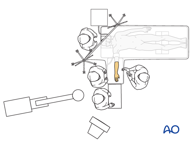
Supine for lateral access
1. Introduction
This position and approach provide good access to the lateral proximal forearm, but medial access is quite limited. Repair of some olecranon and coronoid fractures may be difficult. Consider a posterior skin incision allowing a more extensile approach, with the patient prone or lateral decubitus.
2. Preoperative preparation
Operating room personnel need to know and confirm:
- Site and side of fracture
- Type of operation planned
- Ensure that operative site has been marked by the surgeon
- Conditions of the soft tissues (fracture open or closed)
- Implants, instrumentation, and equipment required
- Patient positioning
- Details of the patient (including a signed consent form and appropriate antibiotic and thromboprophylaxis)
- Comorbidities, including allergies
3. Anesthesia
The procedure is performed with the patient under general anesthesia.
While technically possible, regional anesthesia is not advised as the procedure can be prolonged.
4. Positioning
Place the patient supine with the shoulder abducted and the arm positioned on a radiolucent hand table. The elbow is flexed about 90° or as close as the arm rest permits without the hand hanging over its edge.
Make sure the shoulder is well supported and is not retracted too much posteriorly as this could cause stretch of the neurovascular structures.
Take care to avoid traction on the brachial plexus by not over abducting or extending the arm at the shoulder. To prevent this, the hand table must be at the same level as the operating table.
Use of a sterile tourniquet is determined by surgeon’s preference.
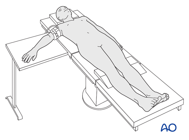
Alternatively, if the arm table is not too large the arm can be placed over the chest as well. This way you can alternate between a lateral approach with the arm on the arm table and a posterior approach with the arm draped over the chest using a single posterior skin incision.
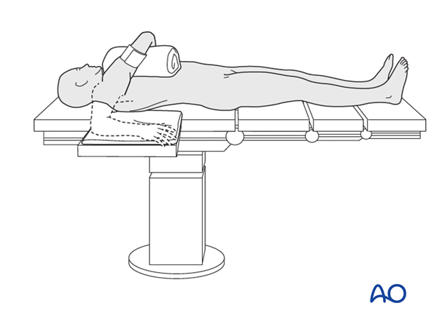
5. Skin disinfecting and draping
After positioning the patient, disinfect the entire upper limb with the appropriate antiseptic.
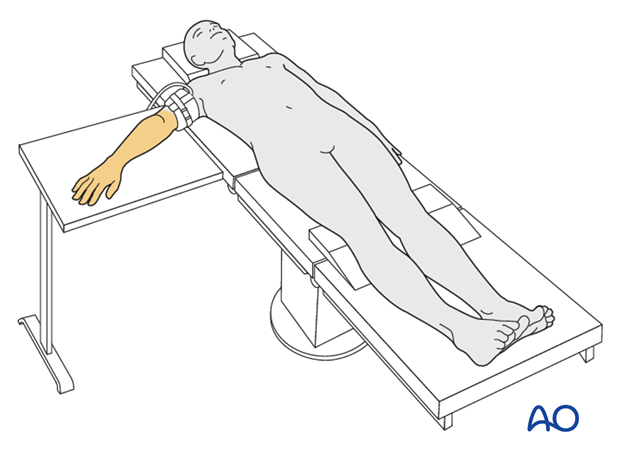
Drape the upper arm, including the tourniquet. If a sterile tourniquet is used it is applied after draping.
Drape the hand separately to allow elbow flexion during surgery and imaging.
Complete patient draping using single-use drapes or sterile sheets.
Drape the image intensifier.
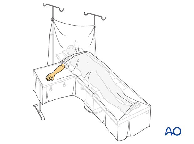
6. Operating room set-up
- The surgeon sits next to the patient’s trunk.
- The assistant sits on the opposite side of the hand table.
- The ORP is positioned next to the assistant.
- Make sure the assistant and scrub nurse do not impede the access of the image intensifier.
- Place the image intensifier display screen in full view of the surgical team and the radiographer.
