Percutaneous reduction techniques (femoral neck fractures)
1. General considerations
It is important to radiographically assess the reduction quality correctly and repeatedly (to recognize intraoperative redisplacement).
2. Reduction techniques
Reduction with a Schanz screw as joystick
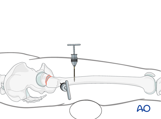
Create a stab incision and insert a Schanz screw in the distal region of the greater trochanter.
Optionally, a second Schanz screw may be added perpendicular in the femoral shaft to better control reduction.
With slight longitudinal traction, reduce any shortening.
If the patient is on a radiolucent table, add a towel or other support under the thigh to avoid posterior translation of the distal fragment.
Internally rotate the distal fragment with the Schanz screw to reduce the fracture.
Check the reduction quality in both views.
Reduction with a bone hook
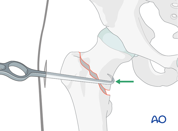
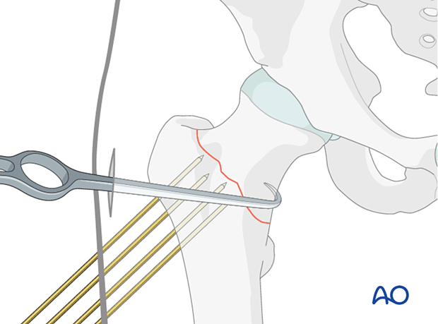
A bone hook can be used to correct valgus angulation.
Make an anterolateral incision at the level of the tip of the greater trochanter to introduce the hook. Carefully insert the hook along the anterior cortex and move it medially around to contact the medial cortex. Use fluoroscopic guidance to ensure proper positioning of the bone hook.
Reduction with a Kelly forceps (Kelly clamp rotation)
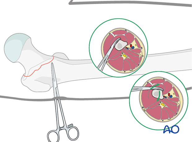
Internal rotation of the distal shaft segment can be achieved with a Kelly clamp introduced percutaneously, which rotates the soft tissue around it.
Insert a Kelly clamp through a posterolateral stab incision. Place its tip on the anterior cortex of the intertrochanteric area.
By lifting the Kelly clamp, the soft tissues and the distal shaft segment are rotated internally.
3. Maintaining reduction
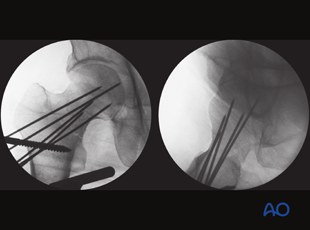
For preliminary fixation, place pins or K-wires through the neck into the femoral head.
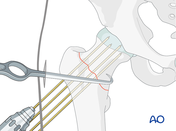
To avoid redisplacement of the fracture by pushing the wires from the lateral cortex into the head, hold the reduction with a hook. This will counteract the lateral force while inserting the K-wires with a power drill.
First, insert multiple K-wires up to the fracture site.
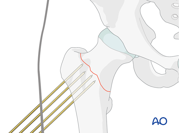
Then pull on the tine of the hook, placed on the medial cortex to counteract the laterally applied forces, ...
... and advance the K-wires with the power drill into the head.
4. Assessment of reduction quality
Assess the reduction quality after provisional fixation in both AP and axial views.
Repeat the assessment of reduction after each step of implant positioning to recognize any intraoperative loss of reduction.
Remove the Schanz screw(s) and/or bone hook.
The K-wires are removed one by one during guide-wire insertion.
An appropriately placed K-wire as seen on the AP and axial views may help select the optimal guide-wire trajectory.
