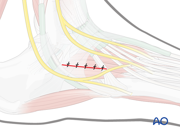
Dorsolateral approach to the midfoot
1. Indications
The dorsolateral approach is indicated for fractures of the lateral column. It is centered over the cuboid and can be extended distally to the base of the 4th metatarsal and proximally to the tip of the fibula.
The following structures can be accessed through this incision:
- 3rd to 5th proximal metatarsals
- 3rd to 5th tarsometatarsal joints
- Lateral cuneiform
- Cuboid
- Calcaneocubiod joint

Combination with the dorsomedial approach
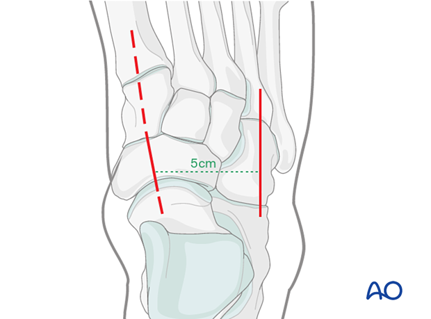
This approach may be combined with the dorsomedial approach. In this case, it is crucial to keep a minimum of 5 cm distance between the two incisions to ensure the survival of the skin bridge.
2. Anatomy
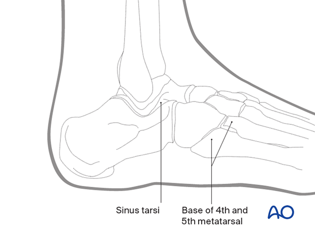
To localize your incision, palpate the following anatomical points.
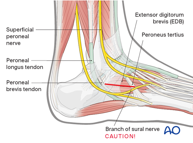
The sinus tarsi and distal calcaneus are palpable proximally. The base of the 4th and 5th metatarsals are palpable distally. The cuboid lies between those two areas.
Key anatomical structures encountered in this approach are the following:
- Extensor digitorum brevis (EDB)
- Peroneus tendons
- Sural nerve
- Superficial peroneal nerve

3. Skin incision
Center the incision over the cuboid along the fourth metatarsal axis and proximally to the tip of the fibula.

Protect the sural nerve
Take care not to harm the dorsolateral or lateral sural nerve branches.
4. Visualization of the joint
In a high-energy injury, the comminution may be severe. Take care not to strip the periosteum or joint capsule from any small pieces. If a piece is attached to a proximal piece of the joint capsule, then the best course of action may be to flip it proximally so as not to disrupt its soft-tissue attachments. Once the joint is reconstructed, this “trap-door” piece can be reduced and fixed.
5. Deep dissection
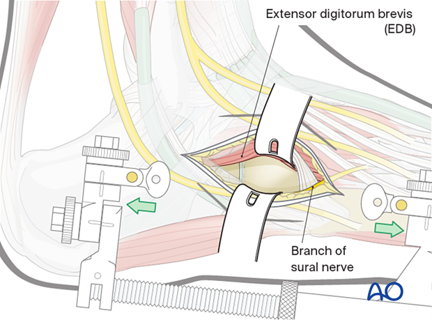
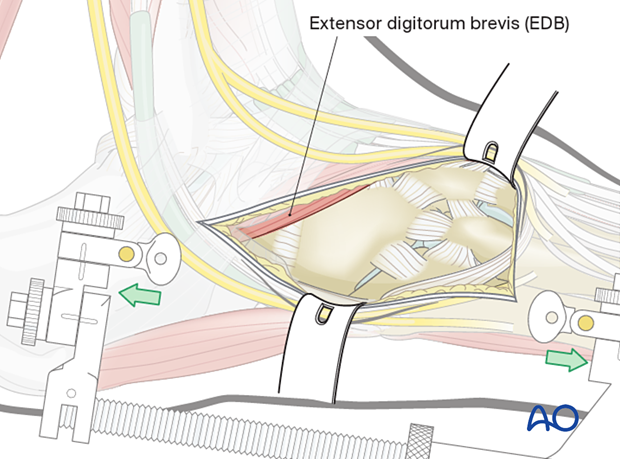
Reflect the extensor digitorum brevis (EDB) muscle belly dorsomedially.
The peroneal muscles can be retracted towards the plantar aspect. The peroneal longus muscle belly crosses from superior laterally and goes under the cuboid in the peroneal groove on its course to the plantar base of the first metatarsal. The peroneus brevis inserts onto the base of the fifth metatarsal.
This exposes the cuboid.

The lateral cuneiform and base of 3rd to 5th metatarsals can be visualized in the intermuscular plane between the extensor digitorum brevis and the peroneal muscles distally.
Closure
The wound is closed in layers taking care to prevent skin tension.
