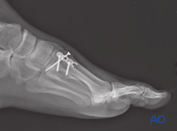
Nonlocking plate with/without lag screws
1. General considerations
Strategy options for complete articular fractures
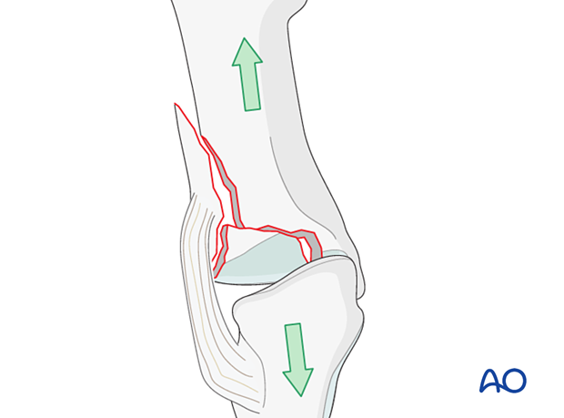
The principle of this procedure is first to reduce and fix the articular block, which is then fixed to the shaft using a plate.
Anatomical reduction of the joint surface is essential to prevent chronic instability or posttraumatic degenerative joint disease.
An alternative strategy is to simultaneously reduce and temporarily fix all components of the fracture, followed by definitive fixation. Typically the definitive fixation would start with screw fixation of the articular block.
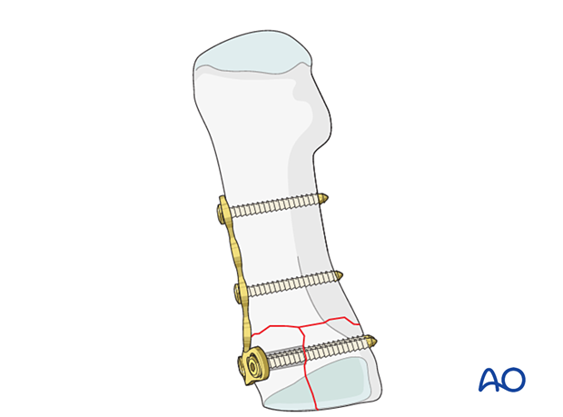
Anatomical considerations
Proper alignment of the metatarsal heads is a critical goal in restoring the forefoot mechanics.
A normal curved “cascade” (Lelièvre’s parabola) appearance, which is symmetric with the other foot, is mandatory on the AP view. See illustration. This symmetry ensures that the normal length of the metatarsal is restored.
It is also critical to restore the metatarsals in their axial or horizontal plane so that all the metatarsal heads are on the same level in the axial view.
Any malalignment, particularly flexion, will recreate focally high pressure during the stance phase and toe-off, resulting in pain and subsequent callus formation.
The sesamoids, rather than the first metatarsal head, bear weight in the first row. Therefore, one must look at the sesamoid level in establishing the alignment in the axial or horizontal plane of the first metatarsal.
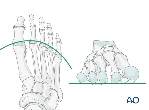
Plate placement on the first metatarsal
Depending on the fracture pattern of the first metatarsal, the plate can be applied medially or dorsally to allow lag screw insertion through the plate, perpendicular to the fracture plane.
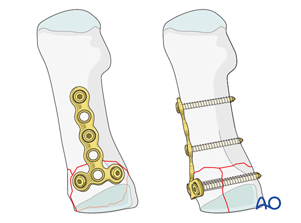
As a dorsal plate will cause less soft-tissue irritation than a medial plate, some surgeons prefer dorsal plate application independently of the fracture patterns direction. This would only be feasible for the illustrated fracture if the articular block is fixed with a medially inserted lag screw (or K-wire), followed by a dorsal plate application. Alternatively, a locking plate would need to be used.
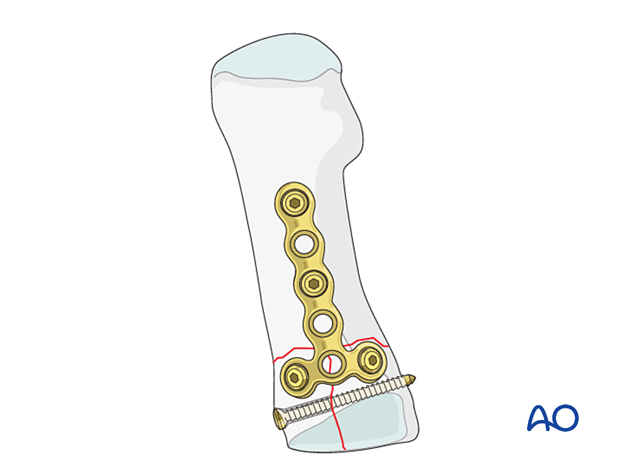
Plate placement on proximal metatarsals 2–4
For metatarsals 2–4, the plate can only be applied dorsally, and consequently, the fracture pattern may not always allow for lag screw fixation.
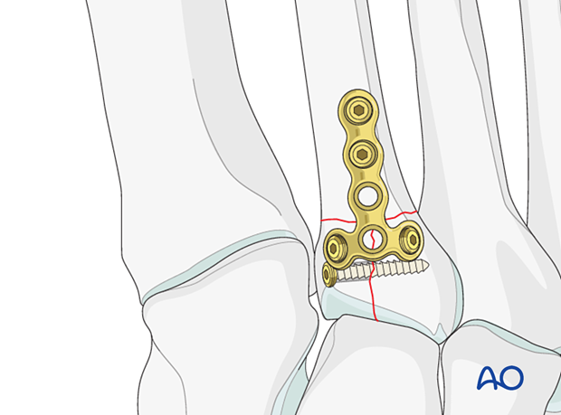
Timing of surgery
The timing of surgery is influenced by the soft tissue injury and the patient's physiologic status.

2. Patient preparation
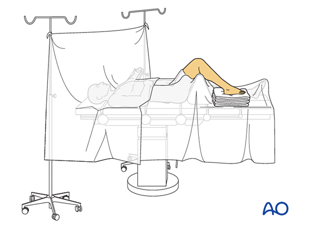
This procedure is typically performed with the patient placed supine with the knee flexed at 90°.
3. Surgical approach
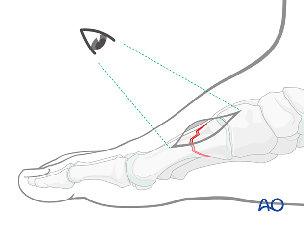
Fractures of the first metatarsal can be approached through a medial or a dorsal approach:
Any single metatarsal injury in metatarsals 2–4 can be approached through the following dorsal approach:
- Dorsal approach to single metatarsals 2–4
- Dorsal approach to multiple metatarsals (when several metatarsals are injured).
4. Reduction
Visualization of the joint
Expose the articular surfaces.
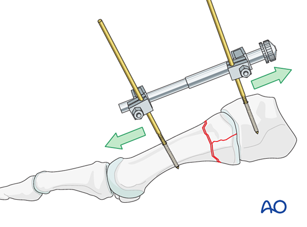
Visualization may be improved by longitudinal manual distraction.
For fractures involving the first metatarsal, a mini distractor may be helpful. Visualization may be improved by releasing the dorsal insertion of the extensor hallucis brevis or extensor digitorum brevis (EHB or EDB).
The extensor hallucis longus should be preserved.
Irrigation
Clean the fracture using a dental pick. Direct suction or irrigation is helpful.
The fracture edges are exposed, and the fragment mobility is assessed. Take great care to avoid further fragmentation. It is essential to maintain the vascularity of small fragments.
The extent of articular involvement is assessed, including separate osteochondral fragments.
If reconstruction of the articular surface is not feasible, a fusion of the joint should be considered.
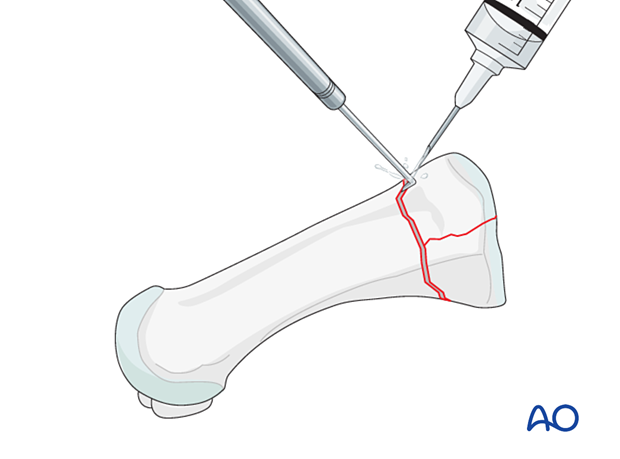
Reduction
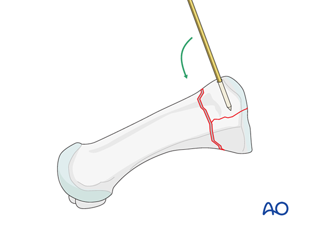
Under direct visualization, the articular fragments are manipulated using a dental pick, small K-wires as joysticks, or a small periosteal elevator.
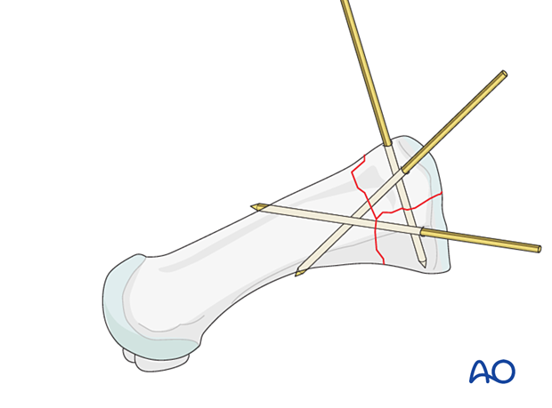
Small K-wires can be inserted across the fracture line for preliminary fixation.
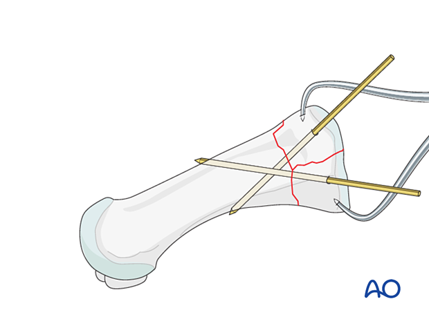
Depending on fracture configuration, pointed reduction forceps may be used for reduction and preliminary fixation.
Be careful not to apply excessive force as this can lead to fragmentation. If possible, apply the reduction forceps so that the forces created by the forces are at right angles to the fracture line. This forceps placement helps in reducing the fracture and in applying compression.
Reduction of impacted fractures
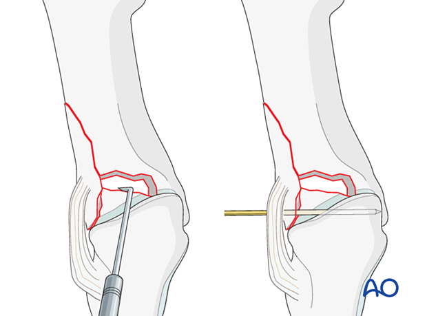
PrincipleCentrally impacted fragments in compression fractures are devoid of soft-tissue attachments and are therefore not reducible by ligamentotaxis.
Direct reduction is necessary.
The key to fixing compression fractures is restoring the joint surface to as close as normal as possible and supporting the reduction with bone graft and internal fixation.
Using a K-wire, or dental pick, the fragments are disimpacted and pushed towards the opposing bone, which is used as a template to ensure congruity of the articular surface.
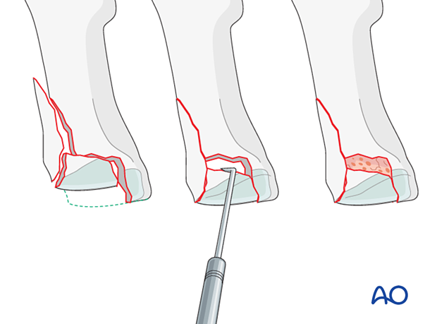
Since the metaphyseal cancellous bone is impacted, a void may be created by its disimpaction.
This void jeopardizes the fracture in two ways:
- It is a very unstable situation in which the articular fragments may easily displace (collapse)
- The healing process is delayed
The bony defect under the fragments is filled with cancellous bone graft. The bone graft also helps keep the fragments in place when the screw fixation is performed.
Visual inspection of the reduction
Anatomical reduction of the joint surface must be checked under direct visualization and image intensification.
Length, alignment, and rotation of all metatarsals and joints must be maintained.
5. Fixation of the articular block
Plate selection and contouring
A 2.0 or 2.4 T-plate is typically used. The length of the plate should allow at least two points of fixation in the shaft.
Contour the plate to fit the bone surface of the metatarsal.
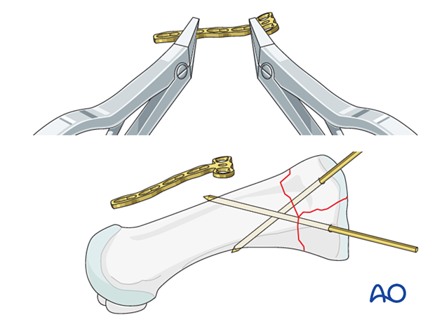
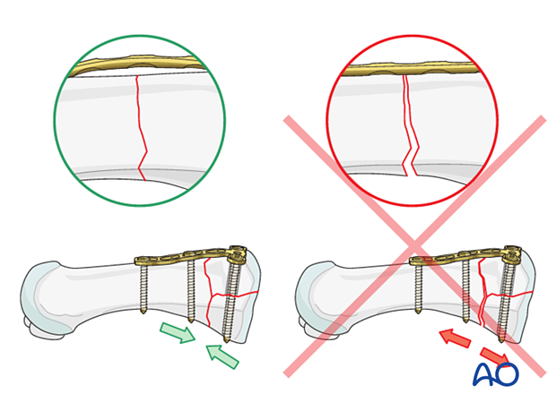
The plate should be slightly over-contoured when applied to a simple extraarticular fracture component. Over contouring will allow fracture compression across the entire extraarticular fracture plane during screw tightening.
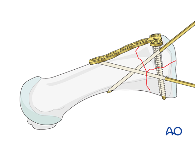
Fixation of the articular block
Fix the articular block using lag screws. The screws can be inserted through the plate if appropriate.
6. Reduction and fixation of the articular block to the shaft
Reduction (if not already reduced)
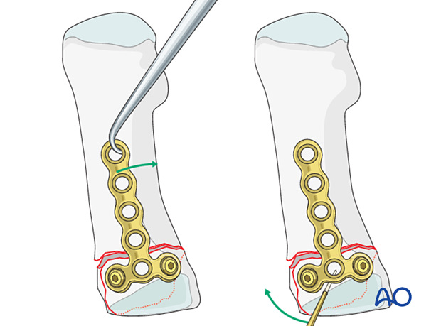
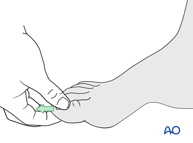
Reduce the articular block to the shaft and restore anatomical axial rotation, length, and angulation.
A periosteal elevator used as a lever may be helpful.
Alternatively, use a reduction forceps, dental pick, or a K-wire as a joystick as described above.
If there is any comminution in the shaft component, the fracture will shorten.
The length can be restored either by manual traction or a distractor.
A distractor will also maintain the length, whereas a manual distraction will not.
Verification of reduction
Use image intensification to confirm the reduction.
Fixation
Simple shaft fracturesThe first screw should be inserted in compression mode if a compression plate is used. If further compression is desired, the second screw can also be inserted in compression mode.
The articular block is fixed to the shaft by inserting two 2.0 mm or 2.4 mm screws inserted in neutral mode.
In this illustration, a compression plate is not used. Here a small clamp is used to stabilize the fracture. Use a small bone hook to create compression at the fracture site before screw insertion.
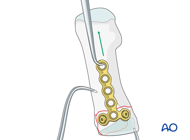
The articular block is fixed to the shaft by inserting two 2.0 mm or 2.4 mm screws without compression.
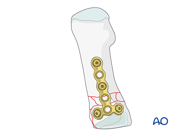
7. X-rays
Obtain x-rays to confirm length, rotation, angulation, and hardware placement.
8. Aftercare
An appropriate well-padded dressing should be applied to protect the surgical incision. Compression will help control swelling.
If present, the skin-pin interface should be similarly well-padded but with dressings that can be readily removed to inspect for pin site infection.
Immediate postoperative treatment is rest, ice, and elevation.
The patient should restrict weight-bearing for six weeks until signs of radiographic healing are present. After this, patients can be weight-bearing as tolerated.
Patients must exercise their ankle and subtalar joints out of the orthosis to prevent stiffness (eg, by stretching their Achilles).
X-ray the metatarsals at six weeks to confirm satisfactory union and remove K-wires if present. Once the fracture is united, the orthosis may be gradually discontinued.
A gastrocnemius release may need to be performed in cases with postoperative gastrocnemius contracture. This occurs more typically in the mid- and hindfoot.
If the gastrocnemius muscle has been released, a splint or cam walker can be used to protect the surgical site.
9. Case 1
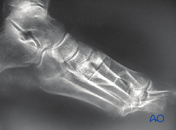
This case shows a significant foot injury consisting of a partial articular fracture of the 1st metatarsal combined with 2nd -4th metatarsal fractures.
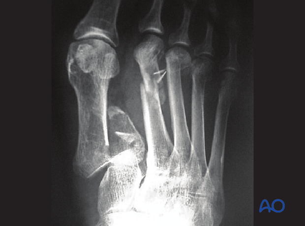
Lateral image of the same injury
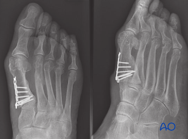
AP and oblique image showing reduction of the first metatarsal fracture with resultant alignment of the 2nd – 4th metatarsal fractures.
Often the first metatarsal is the key to the reduction of the other metatarsals because it bears much more biomechanical stress.
The fixation of the 1st metatarsal with subsequent nonoperative management of the 2nd- 4th metatarsal may be a very reasonable choice.
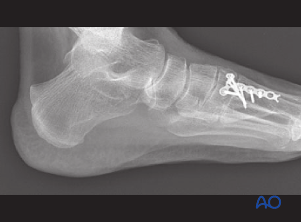
Lateral image.
10. Case 2
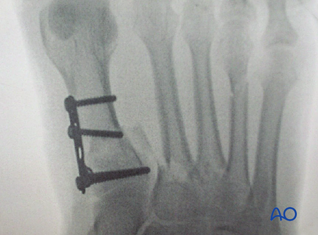
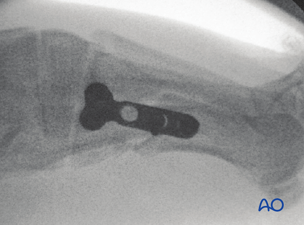
This case shows a significant foot injury with an extraarticular base fracture of the 1st metatarsal with associated fractures of the 2nd–4th metatarsals.
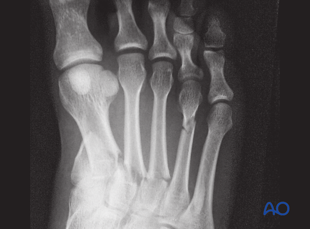
AP image showing reduction of the first metatarsal fracture with the resultant alignment of the 2nd–4th metatarsal fractures.
Often the first metatarsal is the key to reducing the other metatarsals because it bears much more biomechanical stress.
The fixation of the 1st metatarsal with subsequent nonoperative management of the 2nd–4th metatarsal may be a very reasonable choice.
Lateral image
11. Case 3
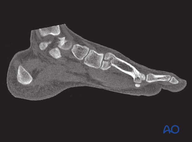
Preoperative CT scan showing a simple displaced articular split fracture of the base of the first metatarsal.
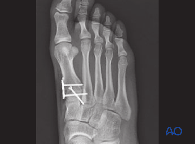
AP image of the postoperative reduction and fixation.
A lag screw was used to fix the articular block, while a mini fragment plate was used to fix the articular block to the shaft.
Lateral view
