Reduction - Immobilization
1. General considerations
Joint dislocation, with or without small bony or ligamentous avulsions, may be reduced closed, and the stability should then be evaluated. Often more than one carpometacarpal (CMC) joint is affected.
If closed reduction is not successful, obstructing soft tissues or fragments need to be removed by exposure of the affected CMC joints.
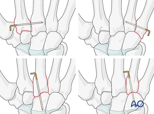
If there is persistent instability, the joint may be stabilized with a temporary K-wire. In noncompliant patients, K-wire stabilization is recommended.
2. Patient preparation
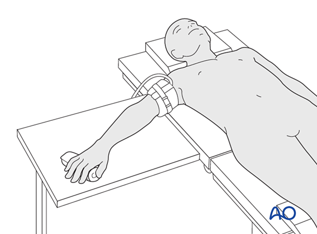
Place the patient supine with the arm on a radiolucent hand table.
3. Approaches
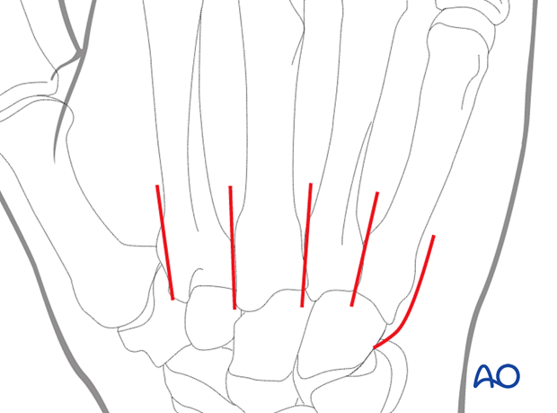
A dorsal approach to the affected CMC joints may be used.
If only the 5th CMC joint needs exposure, an approach to this joint may be used.
Protect the dorsal sensory nerve branches (radial and ulnar).
4. Reduction
Dislocation is usually dorsally and may be reduced manually in a closed manner.
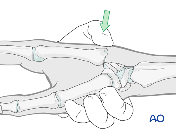
Open reduction is indicated if there are interposed soft tissue or bony fragments. Often, the joint capsules are ruptured, and the joint space is easily exposed.
Remove any soft tissue or bony fragments and reduce the joint.
Stability evaluation
Confirm reduction with an image intensifier and check the joint stability by passive flexion and extension of the fingers.
5. Joint stabilization with temporary K-wire
Often there is persistent instability, eg, subluxation or redislocation during the range of motion. In this case, add a temporary K-wire:
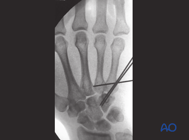
- Transverse fixation of the affected metacarpal base to an uncompromised neighboring metacarpal base
- Retrograde transfixation through the metacarpal base into the carpal bones
Repair the capsule if an open reduction was performed.
Bend the end of the K-wire above the skin and cut it with enough length to avoid migration.

This case shows K-wire stabilization of the 5th to the 4th metacarpal base and the hamate.
6. Aftercare
Postoperative phases
The aftercare can be divided into four phases of healing:
- Inflammatory phase (week 1–3)
- Early repair phase (week 4–6)
- Late repair and early tissue remodeling phase (week 7–12)
- Remodeling and reintegration phase (week 13 onwards)
Full details on each phase can be found here.
Postoperative treatment
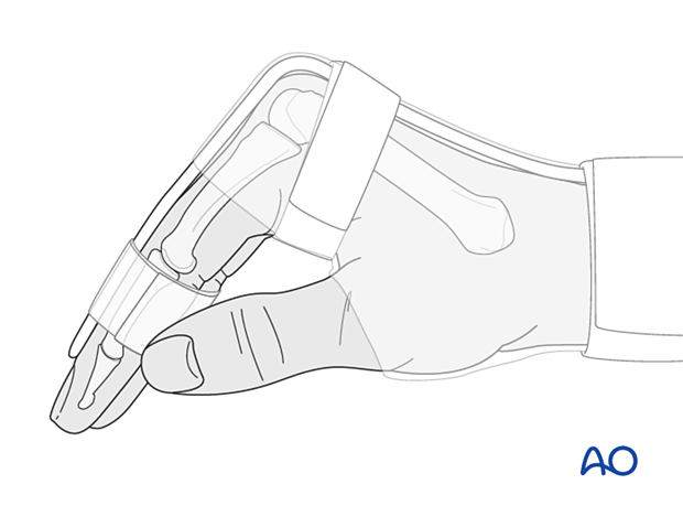
Support the hand with a dorsal splint for about 2–4 weeks. This would allow for finger movement and help with pain and edema control. The arm should be actively elevated to help reduce the swelling.
Pain control
To facilitate rehabilitation, it is important to control the postoperative pain adequately.
- Management of swelling
- Appropriate splintage
- Appropriate oral analgesia
- Careful consideration of peripheral nerve blockade
Follow-up
X-ray checks of joint position have to be performed immediately after the splint has been applied.
Follow-up x-rays with the splint should be taken after 1 week and possibly every 2 weeks.
The K-wire can be removed 4–6 weeks after surgery.
Splint immobilization is continued until about 4 weeks after the injury. At that time, an x-ray without the splint is taken to confirm healing, and range of motion should be pain-free.
Mobilization
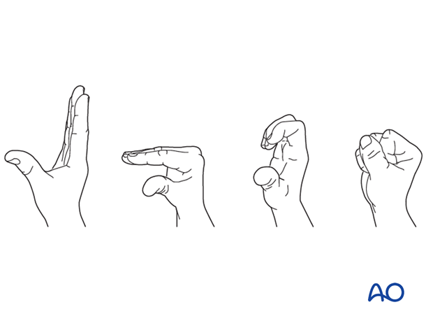
Splinting can then usually be discontinued, and active mobilization is initiated. Functional exercises are recommended.
7. Case
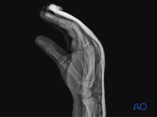
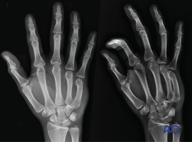
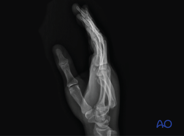
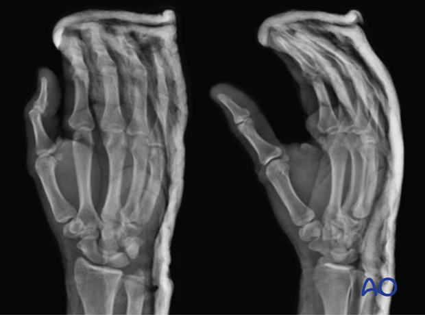
This case shows a dislocated 5th CMC joint.
In the lateral x-ray, the dislocation is seen most prominently.
AP and oblique x-rays showing the hand in a splint with the CMC joint reduced
Lateral view
