Clinical and radiographic examination
1. Introduction
In the AO documentation (1980–1996) 10–14% of all fractures recorded occurred in the forearm.
Simple and wedge fractures are often the result of indirect mechanisms, such as a fall onto an outstretched hand, sporting activities, etc.
Multifragmentary fractures are rarer and are the result of higher energy trauma, such as motor vehicle accidents, falls from heights, high velocity gunshot injuries, etc. The latter are associated with significant soft-tissue damage and should be given high priority.
2. Clinical examination
Patients should be checked for:
- Circulation (radial and ulnar pulses, and also hand perfusion)
- Peripheral neurological status
- Soft-tissue lesions, such as abrasions, lacerations, blisters, skin pathology, and compartment syndrome
3. Radiographic examination
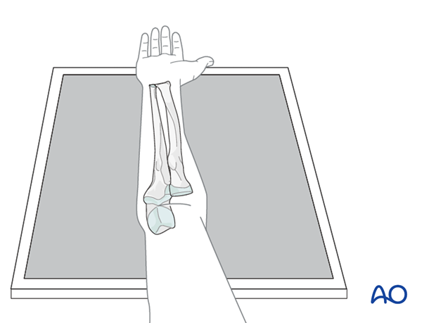
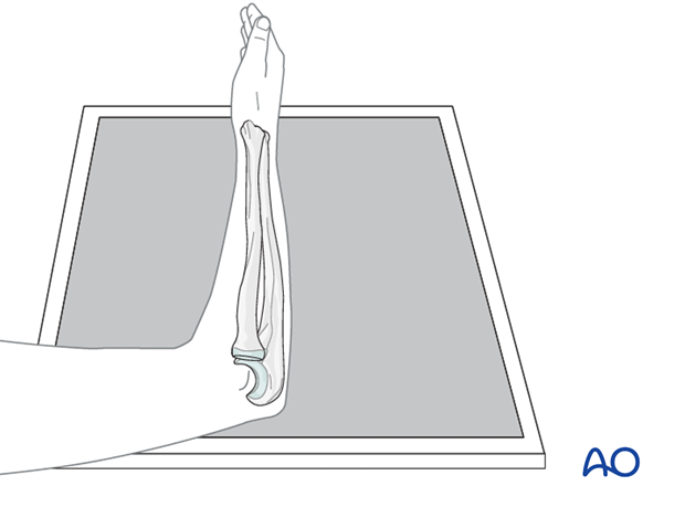
Conventional x-rays of the forearm in two planes are usually sufficient. They must include the elbow and the wrist (but not necessarily on the same image), in order to exclude associated articular injuries and specific fracture patterns, such as Monteggia, Galeazzi, or Essex-Lopresti type injuries.
Correct projections and patient positioning are crucial, particularly, in order to rule out Galeazzi, or Monteggia, fracture-dislocations.
AP view
A sign of a correct projection is a clear view into the distal radioulnar joint (DRUJ).

Lateral view
The projections of the distal and proximal radioulnar joints should each be a true lateral centered on the respective joint.

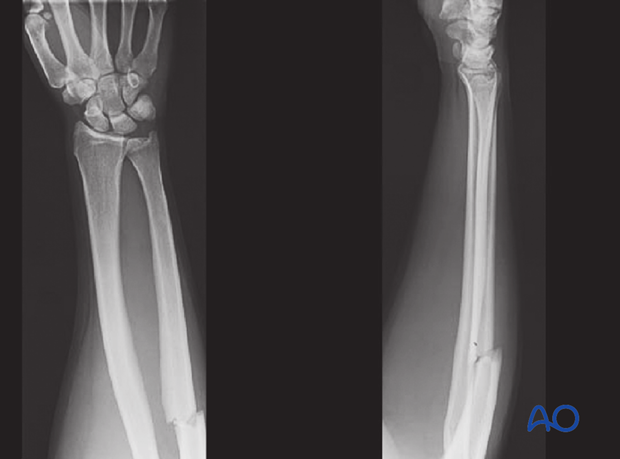
In displaced radial fractures, imaging of the DRUJ is imperative.
These x-rays show good AP and lateral views of the forearm bones with the DRUJ included.

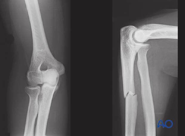
In displaced ulnar fractures, imaging of the elbow joint is crucial.
These x-rays show good AP and lateral views of the proximal forearm bones with the elbow included.

CT scan, or MRI, is rarely required.
In rare cases, when bony anatomy can not be restored, such as in severe comminution, x-rays of the contralateral forearm can be useful. These will help in planning the restoration of the radial and ulnar lengths, thereby maintaining the correct articular relationships in cases in which anatomical reduction is difficult to achieve.


