Lateral approach to the proximal radius
1. Introduction
The lateral approach can be used to access the radial head.
2. Skin incision
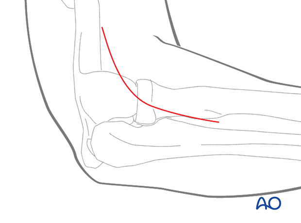
Start the incision 2 cm proximal to the lateral humeral epicondyle.
Carry the incision across the elbow joint, over the radial head, and approx. 5 cm distal to the joint.
Note: The posterior interosseous nerve, within the supinator muscle, crosses the posterior radius, from anteriorly, three finger-breadths distal to the radial head. It must be protected during this approach.

3. Superficial surgical dissection
Incise the subcutaneous tissue and deep fascia in line with the incision.
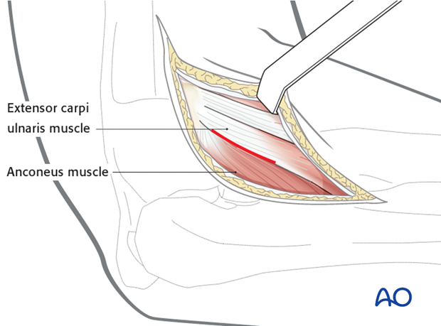
Elevate anterolaterally the subcutaneous tissue and find the interval between the anterior border of the anconeus and the extensor carpi ulnaris muscle.
There may be difficulties in determining the interval between these two muscles because of bruising and bleeding in trauma.

4. Deep surgical dissection
Separate anconeus from extensor carpi ulnaris. Elevate them from the joint capsule.
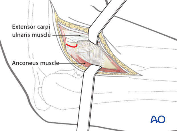
Incise the joint capsule to expose the radial head and the annular ligament.
Note: The annular ligament is entered 1 cm anterior to the ulna to prevent injury to the lateral ulnar collateral ligament.

5. Osteotomy of lateral epicondyle
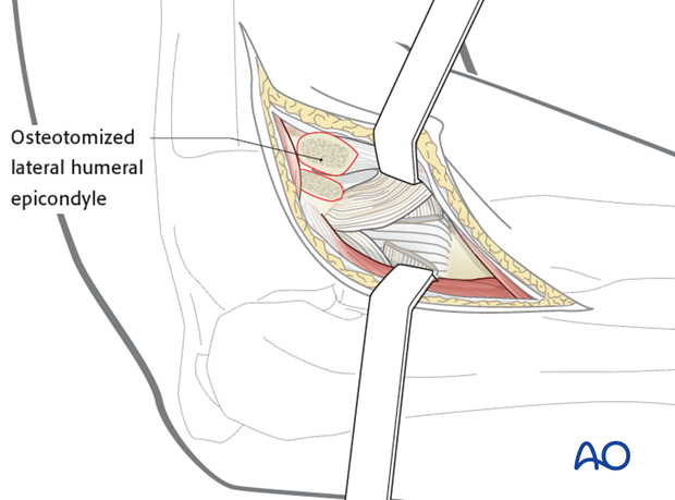
A variation for lateral exposure with preservation of collateral ligaments and extensor tendon origin involves osteotomy of the lateral humeral epicondyle. The osteotomy line in the illustration is marked in red.

The soft tissues and epicondyle are reflected anteriorly to provide access to the proximal radius and ulna.
Repair after this approach requires fixation of the epicondyle. The necessary screw hole can be drilled before the osteotomy.

6. Avoiding damage to radial nerve
- Fully pronating the forearm protects the posterior interosseous nerve by moving it away from the operative field.
- Beware of incising the capsule too far anteriorly as the radial nerve lies over the front of the anterolateral portion of the elbow capsule.
- Beware of dissection distal to the annular ligament or strenuous retraction, because the posterior interosseous nerve lying within the supinator muscle is at risk.


