Approaches to the distal radioulnar joint (posterior/anterior)
1. Indication
Approaches to the distal radioulnar joint (DRUJ) become necessary in cases in which ulnar head dislocations, in the context of Galeazzi injuries, cannot be reduced closed.
In majority of cases, the displacement of the ulnar head is posterior. Very rarely, it dislocates anteriorly.
The approach chosen (whether posterior or anterior) depends on the direction of the displacement.
2. Anatomical consideration
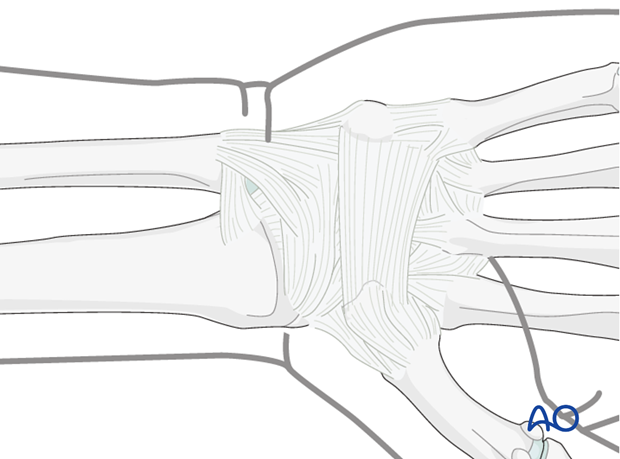
Anatomically, in contrast to the proximal radioulnar joint, there are no clearly defined ligamentous structures in the DRUJ. It is simply a strong joint capsule.
3. Anterior approach
Structures at risk
Be aware of the median nerve to the radial side and the ulnar nerve and vessels to the ulnar side.
Skin incision
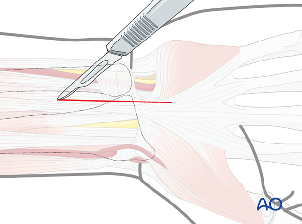
Incise the skin on the medial aspect of the palmaris longus tendon. Be aware of the ulnar nerve and vessels that run along on the lateral aspect of the flexor carpi ulnaris tendon.
Deep dissection
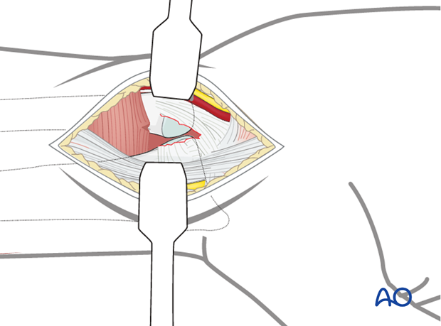
Retract laterally the palmaris longus tendon, median nerve and all flexor tendons. The ulnar nerve and vessels are retracted to the medial side; the distal portion of the pronator quadratus is incised longitudinally, thereby exposing the torn distal articular capsule and the dislocated ulnar head.
Pitfall: Do not retract the median nerve medially, as this would damage the palmar branch of the median nerve.
Repair of articular capsule
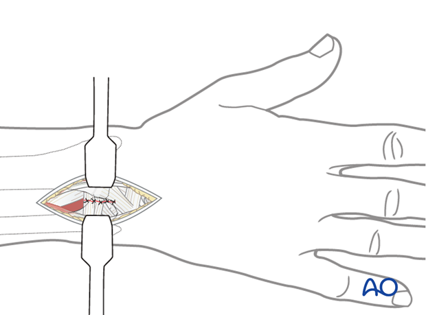
Reduce the ulnar head by pronation and suture the torn ends of the articular capsule using interrupted resorbable sutures.
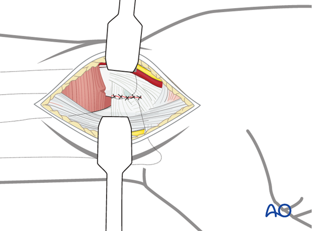
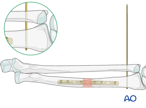
To avoid any redislocation, temporary K-wire transfixation is necessary.
Insert the K-wire(s) in the position of the forearm in which a stable reduction was achieved, i.e., mostly in a neutral or supinated position (as illustrated). The transfixion wire is usually retained for approximately 3 weeks. Most surgeons recommend cast protection during the time of K-wire fixation, to prevent forearm rotation.
4. Posterior approach
Skin incision
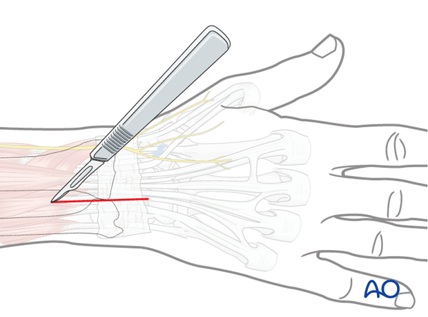
Incise the skin longitudinally in the interval between the ulna and the radius, over the DRUJ.
Deep dissection
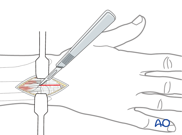
Incise the extensor retinaculum between the 4th and 5th extensor tendon compartments. Retract the extensor digitorum tendons to the lateral side and the extensor tendons to the little finger to the medial side.
Ligate any venous branches of the superficial vein complex.
Repair of articular capsule
Reduce the ulnar head, usually by supination after posterior dislocation, and suture the torn ends of the articular capsule with interrupted stitches of resorbable material. To avoid any redislocation, temporary K-wire transfixation is necessary, with the forearm in the rotational position that led to maximal stability of the DRUJ.
The transfixion wire is usually retained for approximately 3 weeks. Most surgeons recommend cast protection during the time of K-wire fixation, to prevent forearm rotation.
