Complete articular fractures of the proximal hallux
Introduction
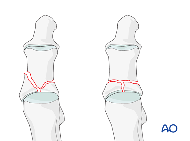
In complete articular fractures, there is complete dissociation of the articular surface from the diaphysis.
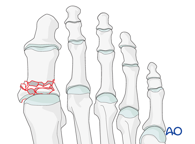
The fracture or the articular surface may be simple or multifragmentary.
Complete articular, simple
Definition
In simple complete articular fractures, there is no comminution of the articular surface. A medial and lateral segment is created. The injury is most commonly associated with joint incongruity with the adjacent joint. Secondary joint congruity is present in certain instances.

Mechanism of the injury
These injuries are usually caused by an axial load to the toe, such as kicking a wall.
Imaging
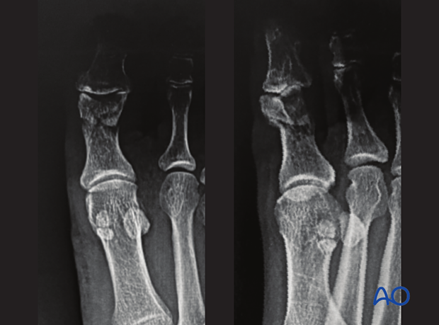
Conventional radiographs of the great toe in AP, lateral, and oblique views are sufficient for diagnosis and treatment.
This case shows a complete articular fracture of the proximal phalanx involving the interphalangeal joint.

Clinical signs
The clinical picture includes swelling, ecchymosis, and pain with range of motion. In some fractures, a deformity may be present.
Complete articular, multifragmentary
Definition
In multifragmentary complete articular fractures, there is usually severe comminution and irregularity of the articular surface. Secondary joint congruity is possible in certain instances.

Mechanism of the injury
Unlike the energy required to generate a simple complete articular fracture, multifragmentary articular fractures derive from high-impact trauma, usually involving axial loading to the toe.
Imaging
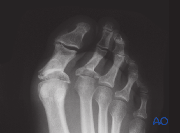
Conventional radiographs of the great toe in AP and lateral oblique views are sufficient for diagnosis and treatment.
The image shows an x-ray of a multifragmentary base of the first and second proximal phalanges.

Clinical signs
The clinical picture includes swelling, ecchymosis, and pain with range of motion. In some fractures, a deformity may be present.


