Nail bed reconstruction
1. Principles
There are three main reasons for nail reinsertion:
- To prevent scarring between the eponychium and the nail matrix.
- To stabilize the fracture.
- The nail acts as a biological barrier and protection.
It is necessary to repair the nail bed or the germinal zone in a precise manner; otherwise, permanent deformity of the nail growth will result.
These procedures are difficult to perform successfully without the help of magnifying loupes.
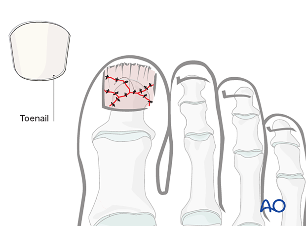
Open fractures
The general principles for treating all open fractures will apply to an open fracture. As most of these injuries are due to crushing, edema of the soft tissues is most likely to develop, and primary closure of any associated skin lacerations is not advisable.
2. Patient preparation
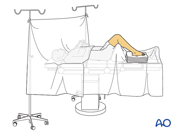
This procedure is typically performed with the patient placed supine and the knee flexed 90°.
This procedure should be performed using a regional block.
3. Release of subungual hematoma
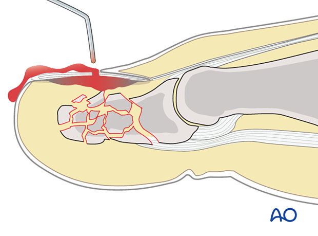
Closed crush injuries are often accompanied by subungual hematoma, which can be exceedingly painful due to the pressure within the closed space.
The hematoma can readily be released by perforating the nail bed with a heated needle.
If no further treatment is indicated, the foot should be splinted, and the hallux protected to avoid any strain due to the motion of the long flexor and extensor tendons.
4. Reduction of displaced bone fragments
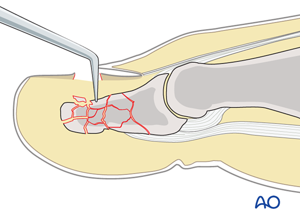
In cases of open fracture, clear the fracture site of blood clots and debris.
Use a dental pick to reduce any displaced fracture fragments carefully. Internal fixation is typically not required.
5. Repair of the nail bed
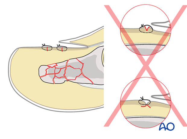
A small-caliber resorbable monofilament is typically used. The sutures can be placed in a simple interrupted manner.
Be careful while suturing the edges of the nail bed. Avoid eversion or inversion to prevent the permanent deformity of nail growth.
6. Reinsertion of the nail
Nail reinsertion
The nail is placed between the eponychium. Then the nail bed and stability of the nail are assessed. If stable, no further procedure is needed.
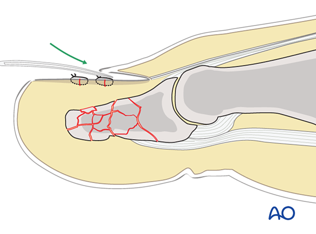
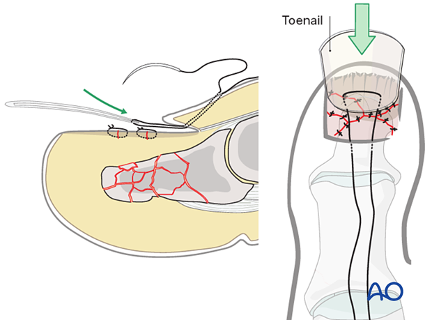
In some patients, the eponychium nail bed interface is injured and prevents it from retaining the nail plate in position.
Begin on the dorsal aspect proximal to the eponychium and insert a suture to exit the undersurface of the eponychium.
Pass the suture through the base of the nail plate as illustrated. The distance between suture passes in the nail should be approximately 5 mm. The suture is then passed through the undersurface of the eponychium to exit through the dorsal soft tissue.
Apply gentle traction on the sutures to reduce the nail plate. Once the nail plate is securely in its bed, tie the suture over a cotton or foam ball to prevent pressure injury to the skin.
Secure distal nail tip
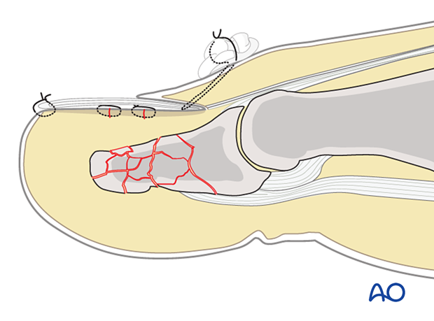
After reinsertion, the nail tends to tilt upwards distally.
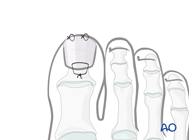
To prevent this, suture the distal nail tip to the nail bed with 2 or 3 interrupted sutures.
Nail loss
In case of nail loss, preserve the nail bed for the growth of the new nail. The nail bed is protected by applying a temporary orthosis made from polyethylene.
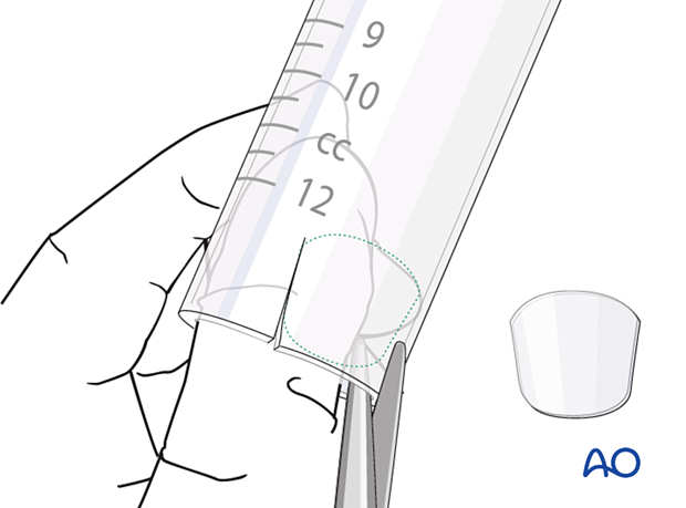
The polyethylene is tailored to the size of the nail...
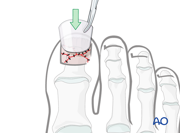
...and then securely fixed with sutures to protect the nail bed using the same technique as nail bed reinsertion.
7. Aftercare
Nail growth will commence, and the new nail will displace the reimplanted nail or polyethylene prosthesis. As the nail grows, the sutures may need to be removed and the prosthesis shortened.
The hallux should be splinted and protected to avoid any strain due to the motion of the long flexor and extensor tendons.
The patient can be weight-bearing as tolerated.


