Distal hallux fractures
Introduction
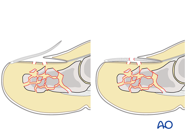
Fractures of the distal phalanx may appear in many different patterns, including impaction fractures of the base with secondary incongruity or multifragmentary fractures secondary to crush, with or without nail bed injuries.
Impaction fracture
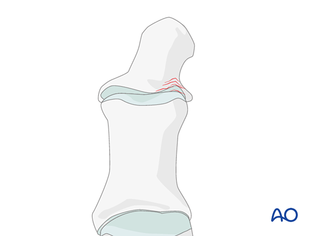
This illustration demonstrates an impaction fracture of the lateral base of the distal phalanx with secondary incongruity.
These patients often present late due to persisting symptoms after a fall or a sprain of the forefoot. A typical example would be an accident with the foot caught under a carpet edge or another obstacle, like a doorstep.
Imaging
Conventional radiographs of the great toe in AP and lateral oblique views are sufficient for diagnosis and treatment.
Simple fracture
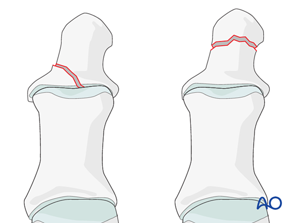
This illustration demonstrates a simple intraarticular and a simple extraarticular fracture.
These patients often present late due to persisting pain and swelling after a fall or a sprain of the forefoot. A typical example would be an accident with the foot caught under a carpet edge or another obstacle, like a doorstep.
Imaging
Conventional radiographs of the great toe in AP and lateral oblique views are sufficient for diagnosis and treatment.
Multifragmentary fractures
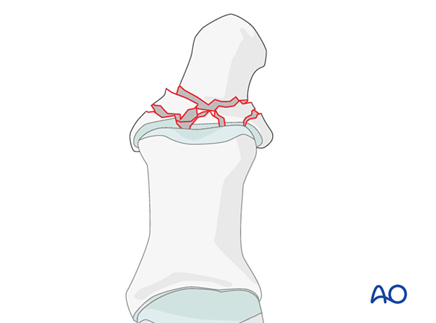
Multifragmentary fractures involve multiple segments of the distal phalanx with or without incongruity of the interphalangeal joint.
These fractures present acutely secondary to obvious trauma. There is immediate pain and difficulty in bearing weight on the affected extremity.
A typical mechanism is the fall of a heavy object directly onto the toe.
Imaging
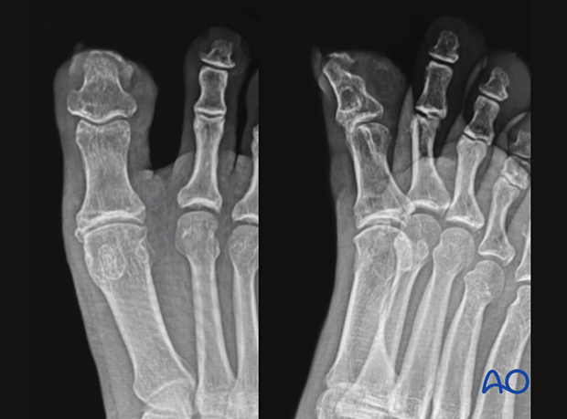
Conventional radiographs of the great toe in AP and lateral oblique views are sufficient for diagnosis and treatment.
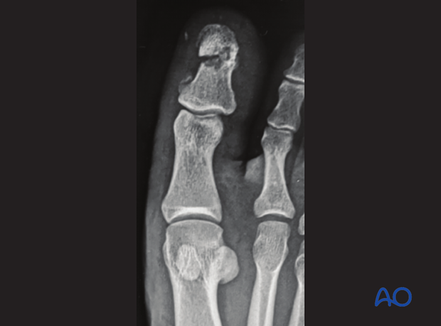
This case shows comminution of the tip of the distal phalanx of the hallux.
Recognizing nail bed injuries
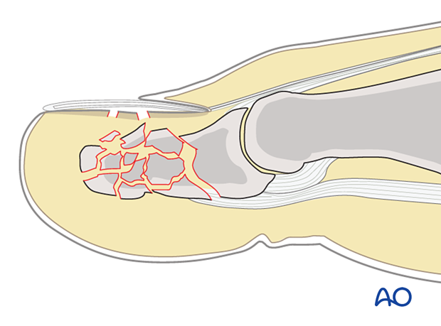
Closed fractures may look harmless on x-rays, but the nail bed has been torn in most cases.
Flexor and extensor tendons displace the fracture with a typical plantar angulation of the tuft fragment.
Subungual hematoma
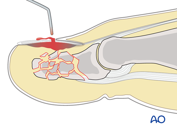
These injuries are commonly associated with a painful subungual hematoma. Non-painful collections do not require evacuations. Painful collections should be expeditiously decompressed with an 18-gauge needle through the nail.
Open fractures
Open fractures present either with an avulsed nail plate or a fractured nail. In both types, the fracture opens dorsally, and the nail bed is also injured.
It is recommended to precisely repair the nail bed.
