Medial approach to the hallux
1. Indications
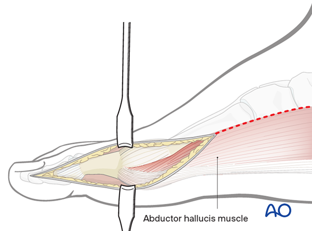
This approach will give access to the medial border of both the proximal and distal hallux.
It is indicated for fractures of the two phalanges of the hallux with or without joint involvement. It can also be used for ORIF of the distal fractures of the first metatarsal. It can also be used to treat hallux rigidus (cheilectomy, osteotomy, fusion).
The incision can be extended proximally to address hallux metatarsal, medial cuneiform, and navicular fractures.
2. Anatomy
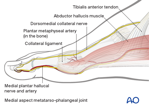
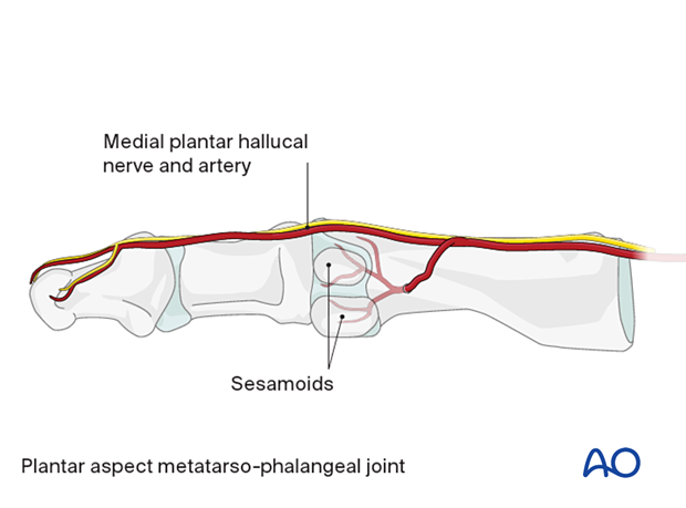
The head of the first metatarsal bone receives its blood supply from an artery, which enters the metatarsal head on the plantar aspect of the distal metaphysis.
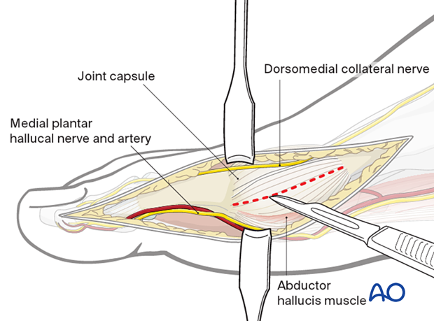
The dorsomedial (collateral) digital nerve (in most cases branch of the deep peroneal nerve) runs on the dorsal half of the medial side, and the medial plantar hallucal nerve runs along its plantar aspect.
The abductor hallucis muscle inserts into the capsule of the MTP joint.
This illustration shows a planter view of the medial plantar hallucal nerve and artery, illustrating the artery's entry point into the bone.
Do not detach the MTP and IP joint's medial collateral ligaments from its attachment site.
3. Skin incision
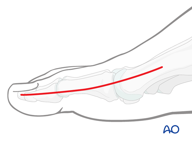
The incision should be centered over the structure to be exposed.
For access to the proximal phalanx, the skin incision starts at the midpoint of the metatarsal head and runs distally towards the mid diaphysis of the distal phalanx.
For access to the distal phalanx, the incision should start at the midpoint of the proximal phalanx and run toward the end of the phalanx, avoiding the nail bed distally.
4. Dissection
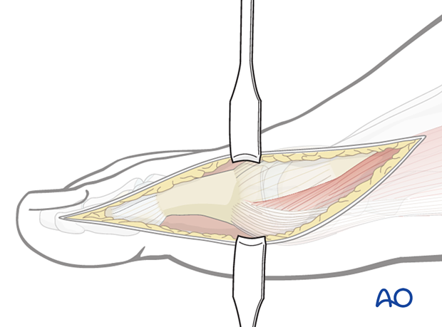
Following the skin incision, dissection is carried through the subdermal tissues directly to the periosteum.
5. Capsulotomy
To expose the proximal or distal articular joint surfaces, incise the capsule in line with its fibers.
Care must be taken to preserve the attachments of the capsule to either side of the joint.

6. Closure
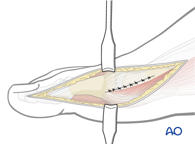
Closure of the capsule
Closure of the capsule can be performed using interrupted sutures.
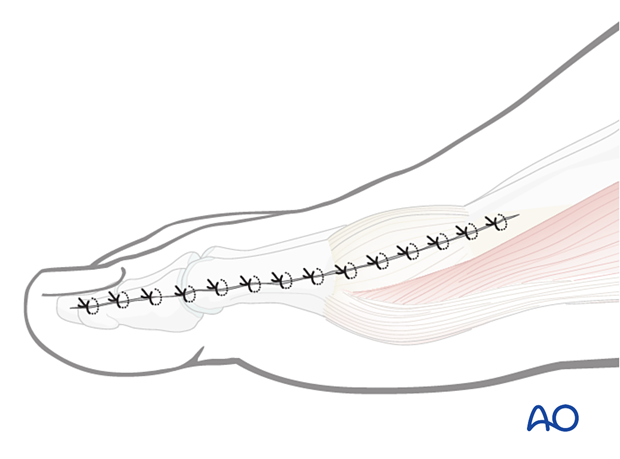
Dermal and skin closure
If possible, the closure is performed in a layered manner, using a resorbable suture in the dermis and nylon or monofilament for the skin.
7. Extension of the approach
One can extend the approach more proximally for diaphyseal fractures of the hallux metatarsal.
