ORIF - Lag screw with protection plate
1. Principles
Lag screw through protection plate
Simple oblique or spiral fractures or intact wedge fractures can be reduced and fixed with lag screws. An additional protection plate is always needed and ideally the lag screw is inserted through the plate.
This procedure can only be carried out as an open one.
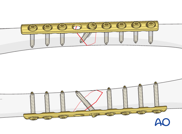
If necessary, the lag screw may be inserted outside the plate.
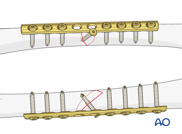
Plate length
It is crucial to use a plate that is long enough so that at least three bicortical screws can be inserted into each main fragment.

2. Patient preparation
The patient may be placed in one of the following positions:
3. Approach
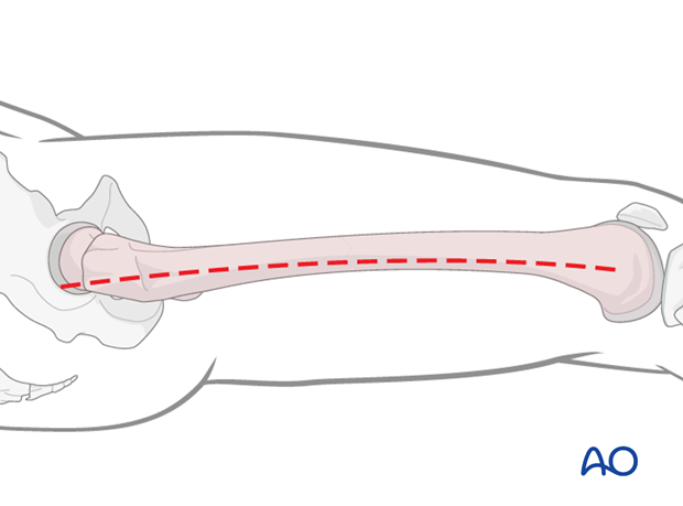
For this procedure a lateral approach is used.
4. Reduction and contouring of the plate
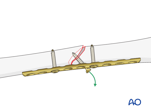
After reduction of the main fragments using pointed forceps, precise contouring of the plate must be undertaken.
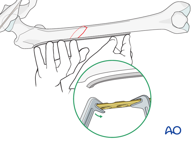
Pitfall: risk of displaced fracture
It is important to contour the plate to fit the bone precisely so that tightening the plate screws does not displace the fracture and might pull out the lag screw.
5. Fixation
Lag screw insertion
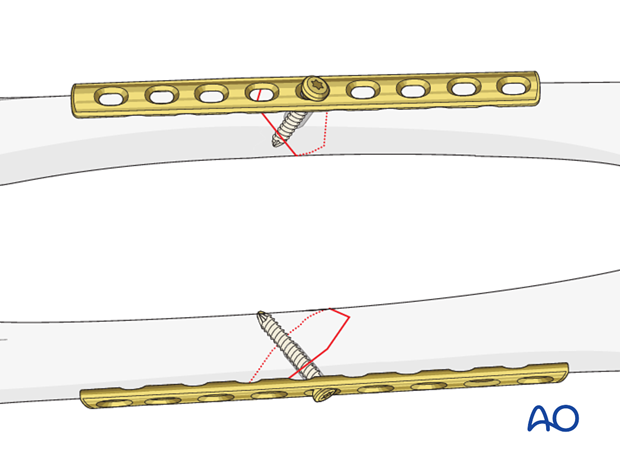
The lag screw is inserted perpendicular to the fracture plane, through the appropriate plate hole. Occasionally, a lag screw of a smaller gauge is used to avoid causing additional fractures.
At this stage, careful handling is mandatory to avoid any stress on the lag screw!
Insertion of bicortical screws
The plate should be fixed to each main fragment with a minimum of three bicortical screws.

Alternative: lag screw outside plate
Illustration shows the completed osteosynthesis with the lag screw inserted outside the plate.

6. Aftercare
Compartment syndrome and nerve injury
Close monitoring of the femoral muscle compartments should be carried out especially during the first 48 hours, in order to rule out compartment syndrome.
Postoperative assessment
In all cases in which radiological control has not been used during the procedure, a check x-ray to determine the correct placement of the implant and fracture reduction should be taken within 24 hours.
Functional treatment
Unless there are other injuries or complications, mobilization may be started on postoperative day 1. Static quadriceps exercises with passive range of motion of the knee should be encouraged. If a continuous passive motion device is used, this must be discontinued at regular intervals for the essential static muscle exercises. Afterwards special emphasis should be placed on active knee and hip movement.
Weight bearing
Full weight bearing may be performed with crutches or a walker.
Follow-up
Wound healing should be assessed regularly within the first two weeks. Subsequently a 6 and 12 week clinical and radiological follow-up is usually made. A longer period may be required if the fracture healing is delayed.
Implant removal
Implant removal is not mandatory and should be discussed with the patient, if there are implant-related symptoms after consolidated fracture healing.


