ORIF - Hook plate
1. Preliminary remarks
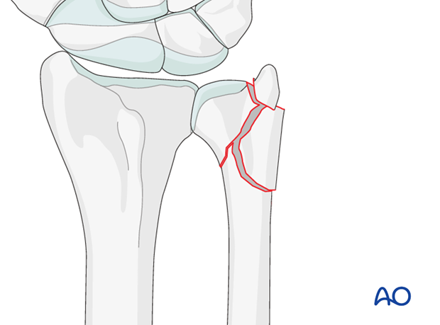
Multifragmentary fractures of the distal ulna usually occur in combination with distal radial fractures.
In multifragmentary ulnar fractures, there is instability and shortening, so the distal ulnar hook plate may be useful to hold distal multifragmentary fractures.
Attention should be paid to restoring correct rotation and correct length in relation to the radius.
Complete dislocation of the radiocarpal joint is often associated with disruption of the distal radioulnar joint (DRUJ).
Teaching video
AO teaching video: Ulna, distal—Subcapital with diaphyseal comminution and styloid fracture. Fixation with a 2.0 LCP Distal Ulna plate
2. Patient preparation and approach
Patient preparation
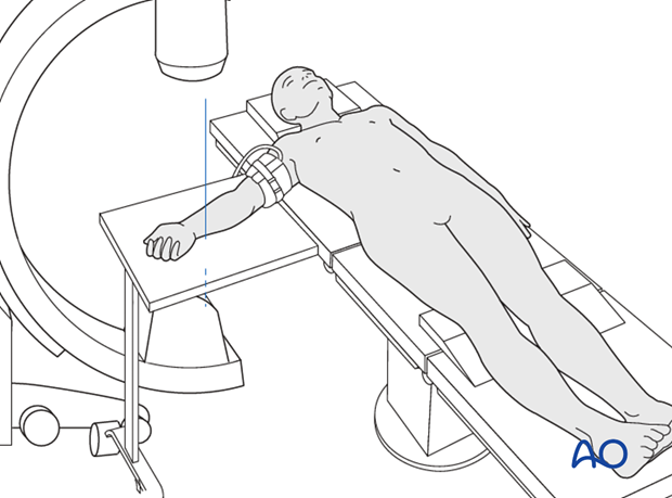
This procedure is normally performed with the patient in a supine position for distal ulnar fractures.
Approach
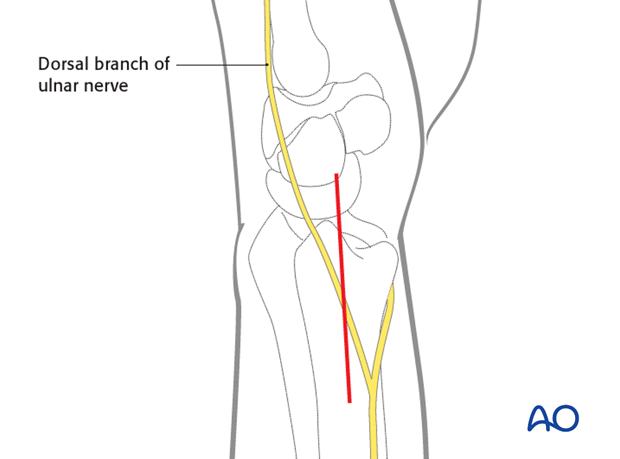
For this procedure an ulnar approach is normally used.
3. Open reduction
Under direct visualization, the ulnar head is reduced to the ulnar shaft using a small periosteal elevator or a dental pick. In multifragmentary subcapital fractures, correct alignment and correct rotational alignment of the head is verified.
A reduction forceps is usually not applicable due to the small fragments and the soft bone quality at this level.
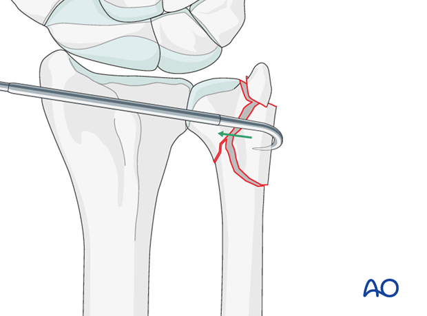
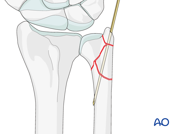
Temporary stabilization with a small K-wire may be helpful, especially if there is a separate ulnar styloid fragment.
4. Internal fixation
Hook plate
The distal ulna hook plate is a precontoured plate that fits to the surface of the distal ulna and allows grasping of the ulnar styloid with the pointed hooks.
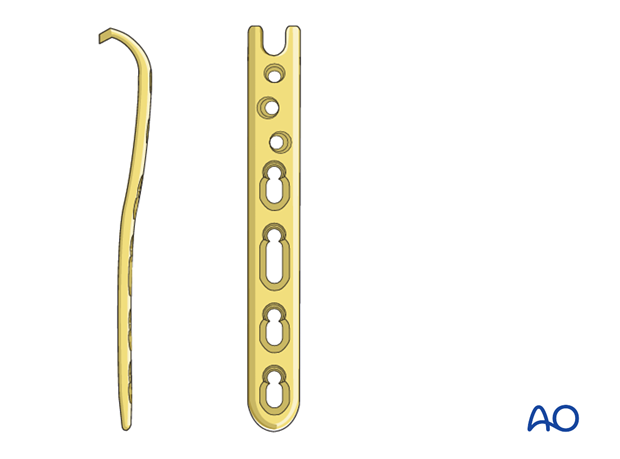
Plate application
The pointed hooks are placed around the tip of the ulnar styloid and the plate is aligned on the ulnar shaft.
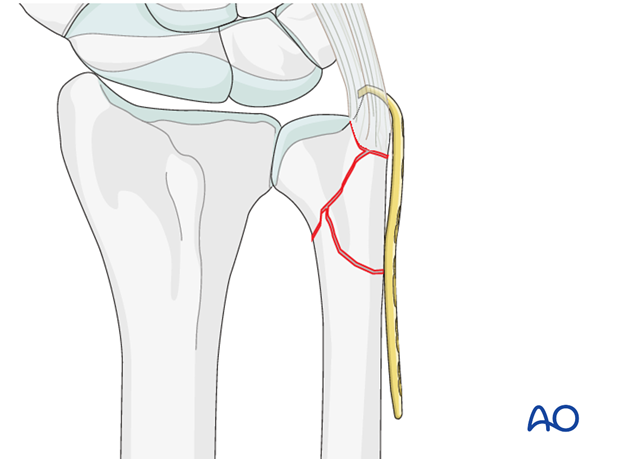
Handling of the plate may be facilitated using the LCP drill sleeve inserted in one of the LCP plate holes.
Image intensification may be used to verify correct plate position.
Screw insertion
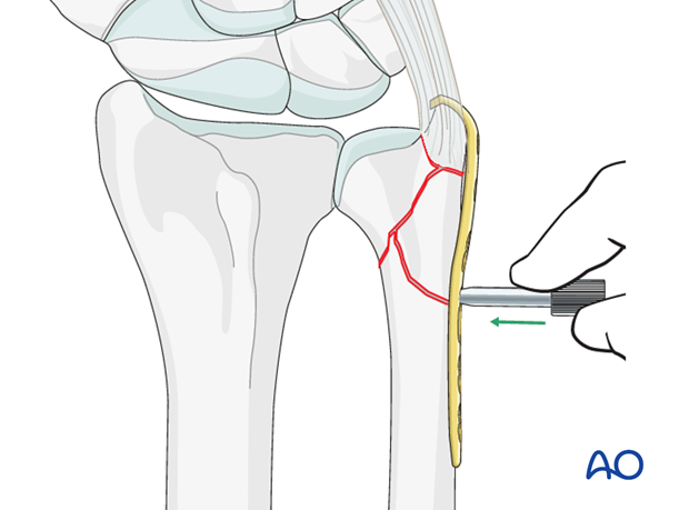
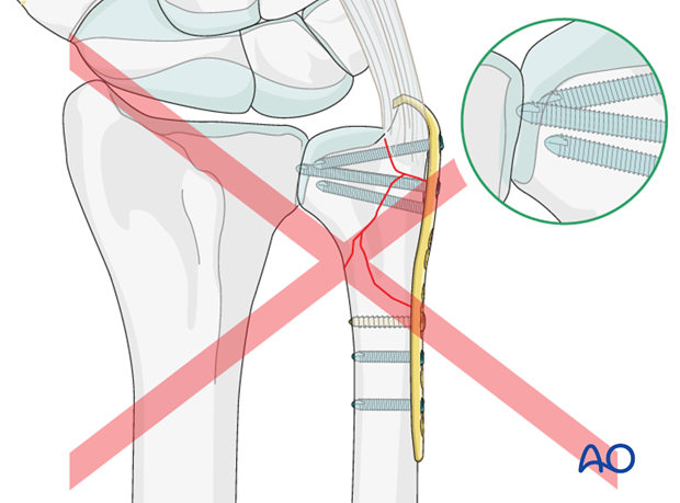
An LCP drill sleeve is used to drill a hole for a locking screw in the ulnar head. Avoid drilling through the opposite cortex, as the screw tip would penetrate the distal radioulnar joint.
Screw length is measured pushing the hook of the depth gauge against the opposite cortex. A slightly shorter screw is then chosen.
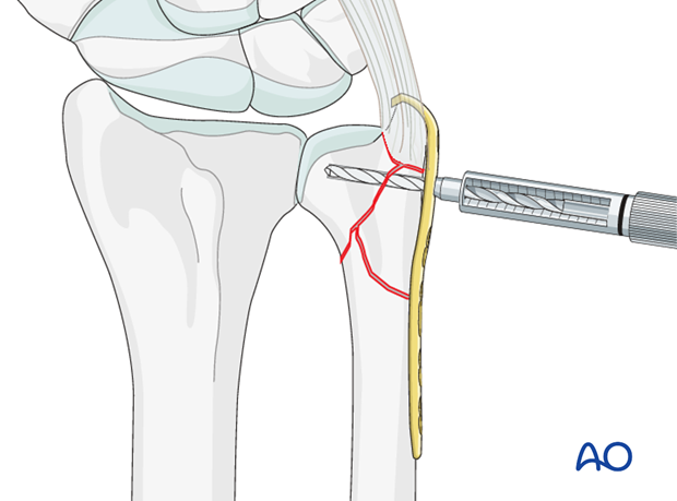
The first locking head screw is inserted into the ulnar head.
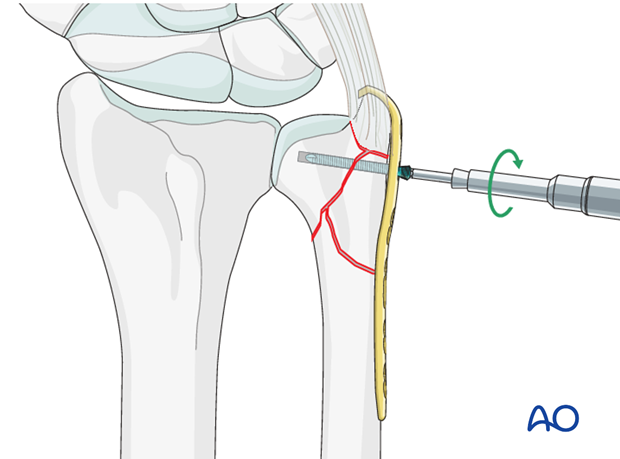
A standard screw is inserted through the oblong plate hole to reduce the shaft fragment to the plate.
At this point reduction is verified under image intensification and unrestricted pronation and supination is checked.
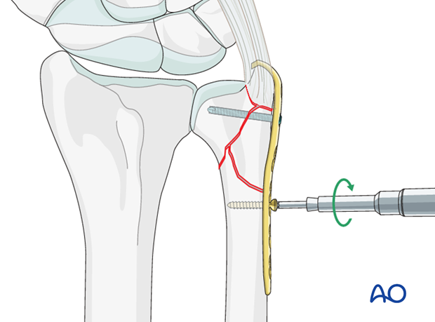
Additional locking head screws are inserted into the ulnar head and fixation at the shaft fragment is completed using standard or locking head screws.
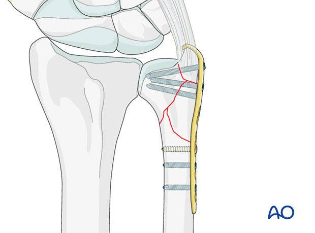
Option
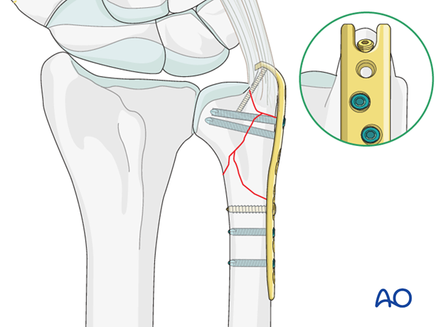
A 1.5 mm screw may be inserted between the pointed hooks of the plate to provide additional stability to the fixation of the ulnar styloid fragment.
In this case, the most distal locking head screw cannot be inserted.
Pearl: Retaining the K-wire
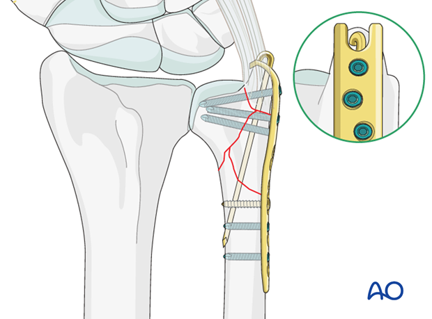
If a K-wire has been used for the fixation of the ulnar styloid, the K- wire may be left in place, if it enters the ulnar styloid between the pointed hooks. The K-wire is then bent and cut short.
Pitfall: First locking head screw too long
If the screw penetrates the opposite cortex of the ulnar head, the screw tip will damage the cartilage of the radioulnar joint.
5. Aftercare
Functional exercises
Immediately postoperatively, the patient should be encouraged to elevate the limb and mobilize the digits, elbow and shoulder.
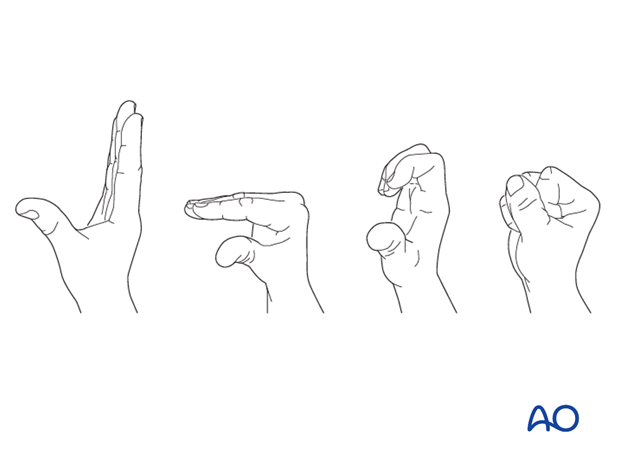
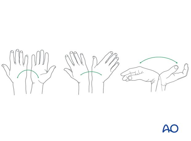
Some surgeons may prefer to immobilize the wrist for 7-10 days before starting active wrist and forearm motion. In those patients, the wrist will remain in the dressing applied at the time of surgery.

Wrist and forearm motion can be initiated when the patient is comfortable and there is no need for immobilization of the wrist after suture removal.
Resisted exercises can be started about 6 weeks after surgery depending on the radiographic appearance.
If necessary, functional exercises can be under the supervision of a hand therapist.
Follow up
See patient 7-10 days after surgery for a wound check and suture removal. X-rays are taken to check the reduction.
Implant removal
Implant removal is purely elective but may be needed in cases of soft-tissue irritation, especially tendon irritation to prevent late rupture. This is particularly a problem with dorsal or radial plates. These plates should be removed between nine and twelve months.


