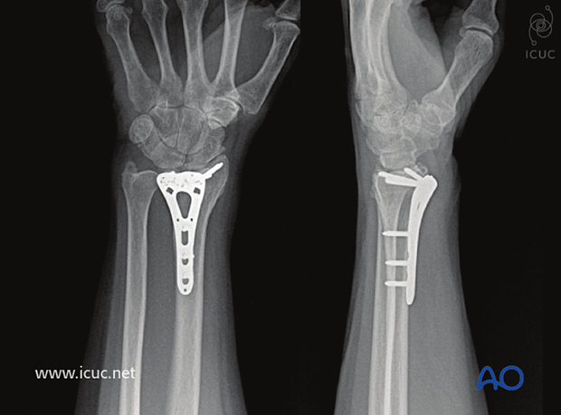
ORIF - Palmar bridge plate
1. Preliminary remarks
Fracture assessment and decision making
These fractures are complete articular fractures of the distal radius.
As these are intraarticular fractures, where possible, they should be treated with anatomic reduction and absolute stability in order to minimize the risk of subsequent degenerative changes in the joint.
Anatomical reduction and stabilization of these articular fractures is also essential because of the functional implications of the involvement of the distal radioulnar joint.
These are among the most common fractures seen and treated in the older population, with underlying osteoporosis. When these fractures occur in younger individuals, they are more likely to be the result of high energy trauma, with associated soft-tissue, or skeletal injuries. It is not possible to make an accurate assessment of the details of these injuries without a CT scan.
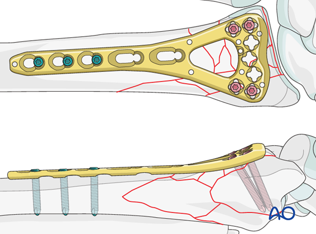
Plate fixation may be applicable for all complete articular fractures, provided the distal fragments are large enough to be held with screws. As long as the articular surface is accurately reduced, and is fixed in the correct position in relation to the radial shaft, it is not necessary to fix all the metaphyseal fragments and the plate may be used in a bridging mode.
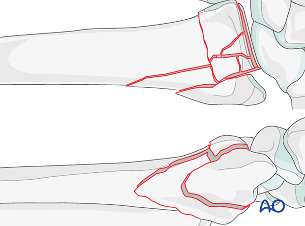
Bridge plating of the distal radius - plate choice
In most complete articular fractures, standard palmar locking plates may be long enough to obtain adequate proximal hold.
When there is comminution involving a significant length of the metaphysis, standard palmar locking plates may not obtain adequate purchase in the proximal fragment, so specially designed, longer angular stable plates have been developed. These provide stable fixation of the distal and proximal fragments, bridging the comminution.
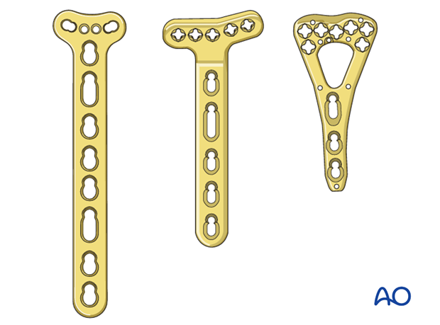
Provisional reduction in displaced fractures
Reduction is achieved by applying longitudinal traction either manually or using Chinese finger traps.
The reduction is maintained by a temporary splint.
If definitive surgery is planned, but cannot be performed within a reasonable time scale, a temporary external fixator may be helpful.
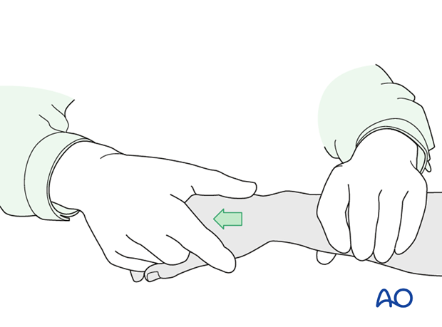
2. Associated injuries
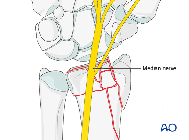
Median nerve compression
If there is dense sensory loss, or other signs of median nerve compression, the median nerve should be decompressed.
Associated carpal injuries
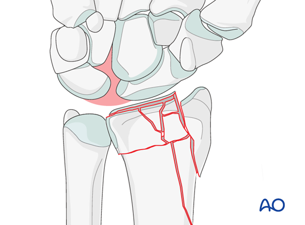
These injuries may be associated with shearing injuries of the articular cartilage, scaphoid fracture and rupture of the scapholunate ligament (SL). Every patient should be assessed for this injury. If present, see carpal bones of the Hand module.
DRUJ/ulnar injuries
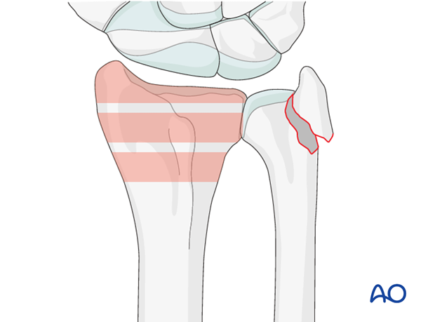
These injuries may be accompanied by avulsion of the ulnar styloid and/or disruption of the DRUJ. If there is gross instability after the fixation of the radial fracture, it is recommended that the styloid and/or the triangular fibrocartilaginous disc (TFC) is reattached. This is not common in simple fractures, but may occur with some high energy injuries.
The uninjured side should be tested as a reference for the injured side.
It may not be possible to assess DRUJ stability until the fracture has been stabilized (as described below).
3. Patient preparation and approach
Patient preparation
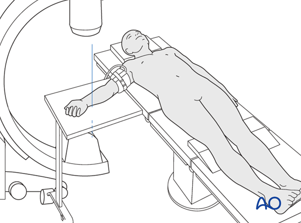
This procedure is normally performed with the patient in a supine position for palmar approaches.
Approach
There are two palmar surgical approaches to the distal radius – a modified Henry approach to the radius and a more ulnar approach, designed to expose the median nerve as well as the distal radius.
A thorough knowledge of the anatomy around the wrist is essential. Read more about the anatomy of the distal forearm.
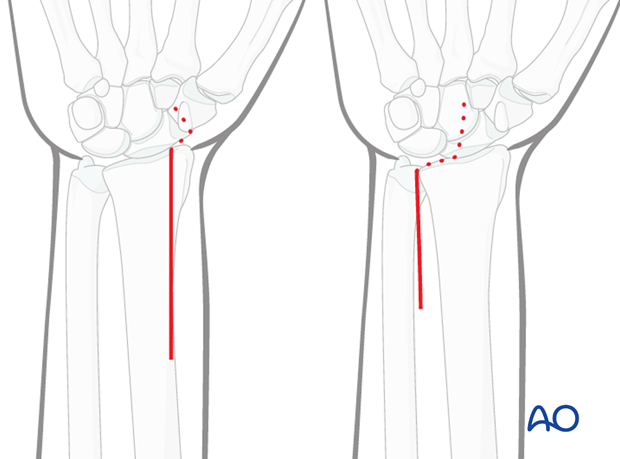
4. Option 1: Reduction with K-Wires and plate fixation
This procedure is used in case of extensive dorsal comminution and intact palmar cortex.
Reduction
Reduce the fracture manually, using longitudinal traction.
Anatomical reduction can be achieved by direct manipulation, using a dental pick or similar hook.
Confirm the reduction under image intensification.
Insert a K-wire across the fracture, through the radial styloid, to provide provisional stabilization.
The major articular fragments may be reduced with the aid of pointed reduction clamp(s).
Temporary fixation with K-Wires of the major articular fragments is an option.
The aim is to achieve as accurate an anatomic reduction of the articular fragments as possible, before the plate is applied.
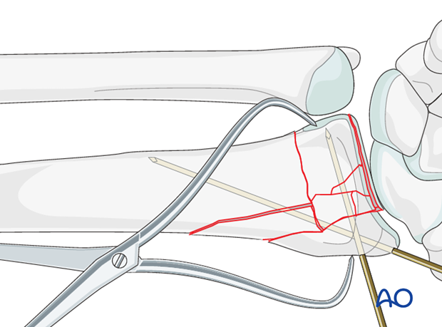
Plate fixation
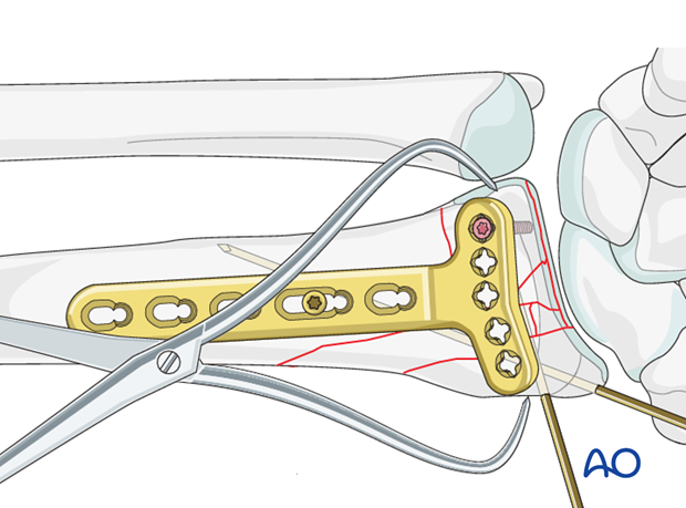
Apply the plate to the bone. The distal end of the plate should end at the anatomic watershed zone of the distal radius.
Insert a screw through the oblong plate hole in the proximal radial fragment. Select a screw which is long enough to engage both cortices.
Insert the screw. Before fully tightening it, check the plate position using intraoperative imaging, adjusting the position of the plate as necessary.
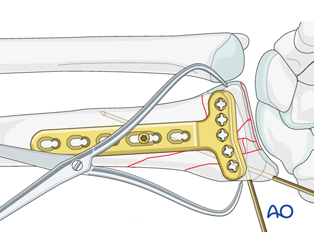
The initial distal screw should be placed through the ulnar sided screw holes.
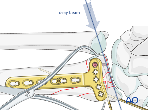
A sagittal image is obtained with the angle of the X-ray beam directed 20° obliquely to the radius to confirm that the screw is not penetrating the radial carpal joint.
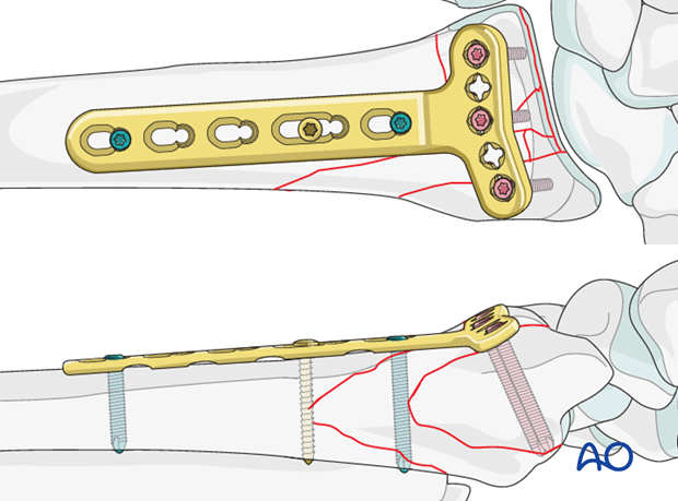
Insert distal locking head screws in a manner similar as described above.
Then insert two more proximal locking head screws.
Remove the K-wires.
5. Option 2: Reduction with spreader/manual traction and plate fixation
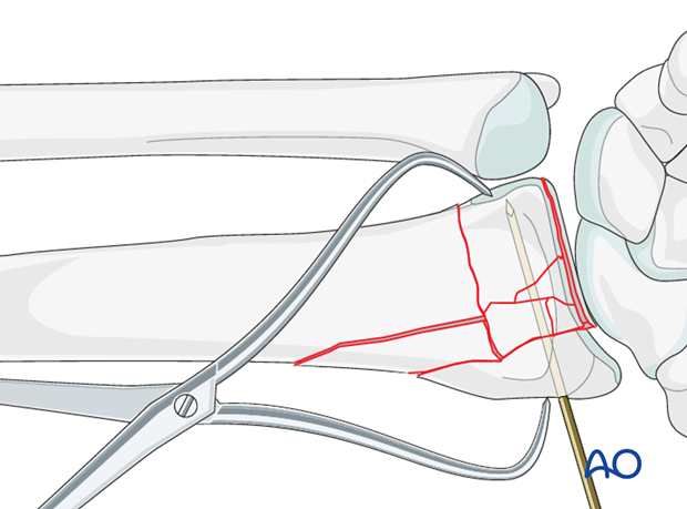
In case of moderate comminution on the dorsal and palmar cortex, it is difficult to maintain temporary reduction with K-wires. So reduction with help of a spreader / manual traction is recommended.
Reduction
The articular block is reduced with the aid of a pointed reduction clamp.
Temporary fixation with K-Wires of the articular block is+ an option.
Radial plate fixation
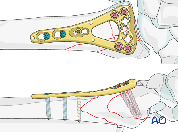
The plate is pre-contoured distally, so it is already adapted to the palmar curvature of the distal radius.
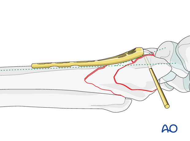
The locking plate is first positioned distally on the flat reduced palmar surface of the distal radius at the anatomic watershed zone and fixed with distal locking screws parallel to the articular surface with at least one, preferably two screw(s) in each articular fragment (depending on the quality of the bone stock).
The first screw inserted is the ulnar one and its position should be checked under image intensification with the hand elevated 20-30° off the table – in lunate facet view.
Once the articular block is securely held to the plate the K-wire(s) may be removed.
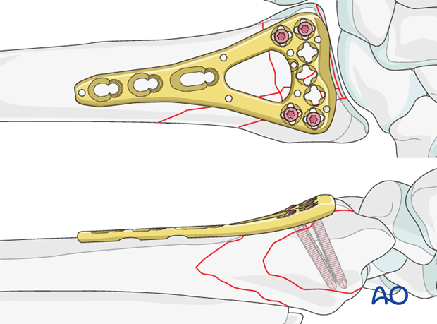
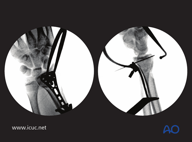
The correct length of the radius in relation to the ulna should be established preoperatively by taking radiograph of the opposite wrist. The length of the radius in relation to the ulna is then achieved by inserting a unicortical screw just proximal to the proximal end of the plate, and then using a spreader, as illustrated, to move the plate gently distally.
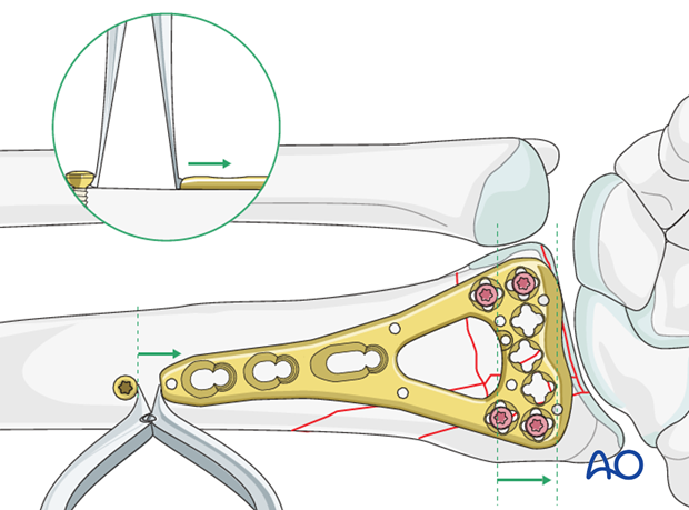
Once the correct length is achieved, the plate is provisionally fixed proximally with one standard screw in the oblong hole.
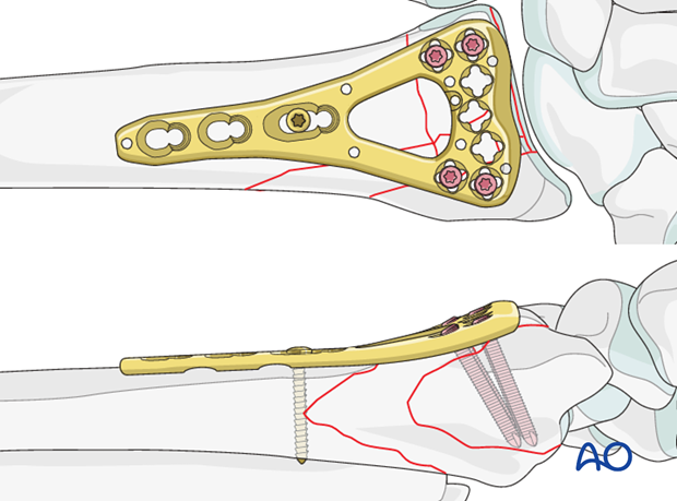
The relationship of the radius to the distal ulna is checked under image intensification before the plate is fixed with additional proximal locking screws.
6. Option 3: Temporary reduction with Ex Fix and plate fixation
In cases of extensive metaphyseal comminution, and/or diaphyseal comminution, reduction may be achieved and maintained with the help of an external fixator.
Reduction
For application of the external fixator, refer to the basic technique for application of modular external fixator.
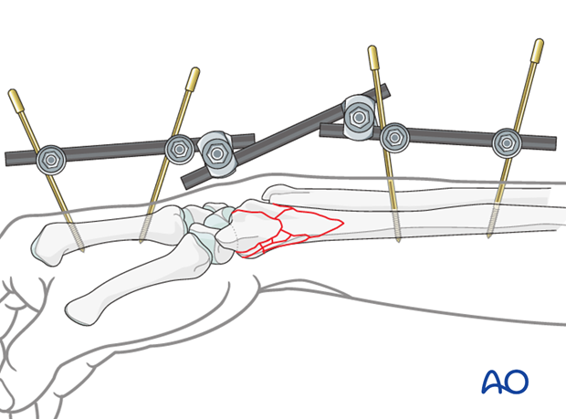
The articular block may be reduced with the aid of a pointed reduction clamp.
Temporary fixation with K-wires of the articular block is an option.
Accurate anatomic reduction of the articular surface must be confirmed before the plate is applied.
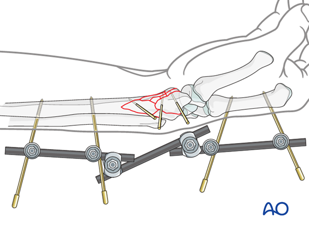
Plate fixation
Once length, axis and rotation of the radius is achieved, the articular surface is reduced, and this is all verified under image intensification, the fracture is plated with an angular stable plate.
Use angularly stable screws for the articular component and the shaft proximal to the comminution to enhance stability.
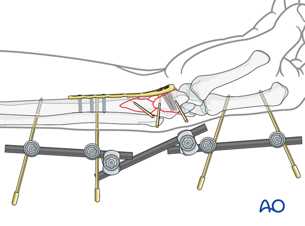
After definitive plate fixation, the external fixator and K-wires are removed.
7. Assessment of Distal Radioulnar Joint (DRUJ)
Before starting the operation, the uninjured side should be tested as a reference for the injured side.
After fixation, the distal radioulnar joint should be assessed for forearm rotation, as well as for stability. The forearm should be rotated completely to make certain there is no anatomical block.
Method 1
The elbow is flexed 90° on the arm table and displacement in dorsal palmar direction is tested in a neutral rotation of the forearm with the wrist in neutral position.
This is repeated with the wrist in radial deviation, which stabilizes the DRUJ, if the ulnar collateral complex (TFCC) is not disrupted.
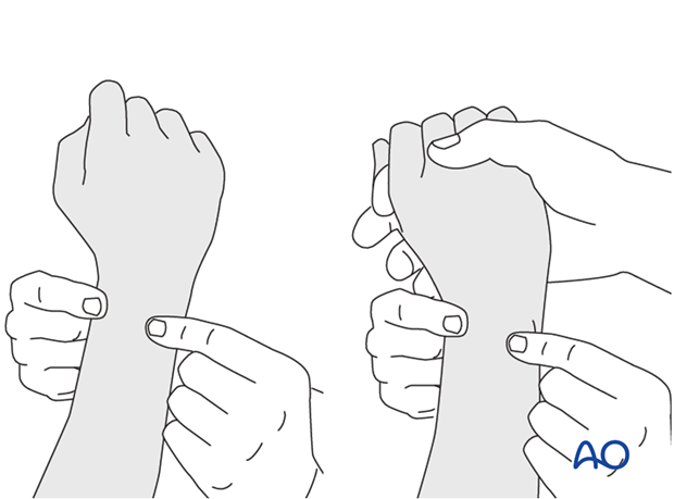
This is repeated with the wrist in full supination and full pronation.
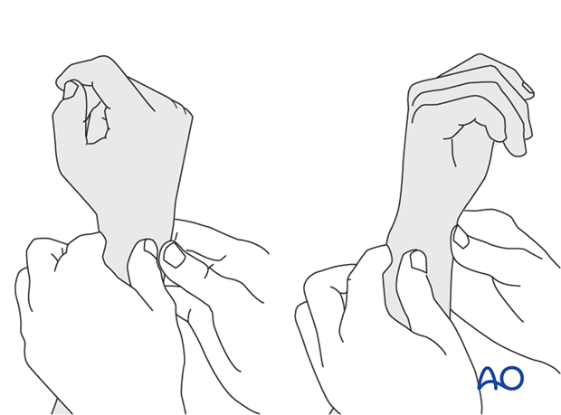
Method 2
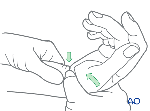
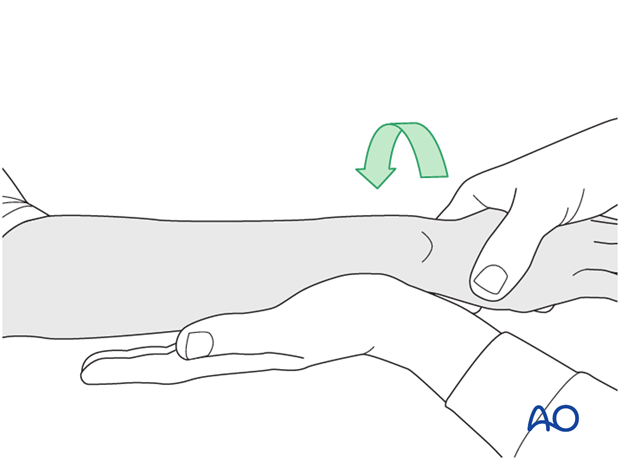
To test the stability of the distal radioulnar joint, the ulna is compressed against the radius...
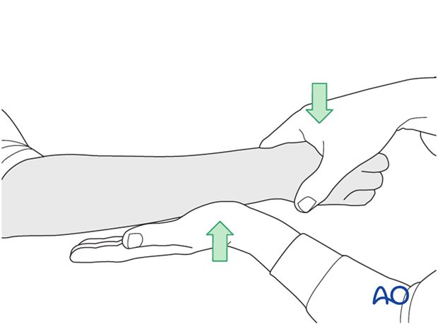
...while the forearm is passively put through full supination...
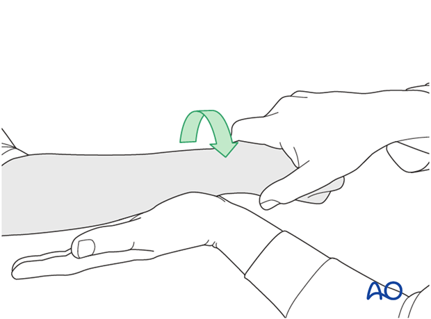
...and pronation.
If there is a palpable “clunk”, then instability of the distal radioulnar joint should be considered. This would be an indication for internal fixation of an ulnar styloid fracture at its base. If the fracture is at the tip of the ulnar styloid consider TFCC stabilization.
8. Aftercare
Functional exercises
Immediately postoperatively, the patient should be encouraged to elevate the limb and mobilize the digits, elbow and shoulder.
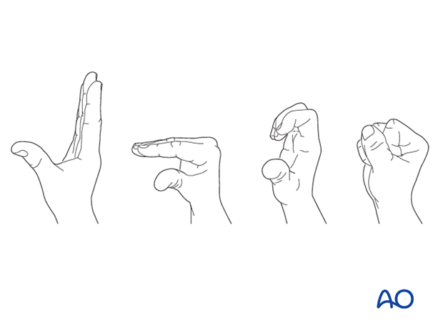
Some surgeons may prefer to immobilize the wrist for 7-10 days before starting active wrist and forearm motion. In those patients, the wrist will remain in the dressing applied at the time of surgery.

Wrist and forearm motion can be initiated when the patient is comfortable and there is no need for immobilization of the wrist after suture removal.
Resisted exercises can be started about 6 weeks after surgery depending on the radiographic appearance.
If necessary, functional exercises can be under the supervision of a hand therapist.

Follow up
See patient 7-10 days after surgery for a wound check and suture removal. X-rays are taken to check the reduction.
Implant removal
Implant removal is purely elective but may be needed in cases of soft-tissue irritation, especially tendon irritation to prevent late rupture. This is particularly a problem with dorsal or radial plates. These plates should be removed between nine and twelve months.
9. Case 1
Fragmentary intraarticular displaced distal radial fracture.
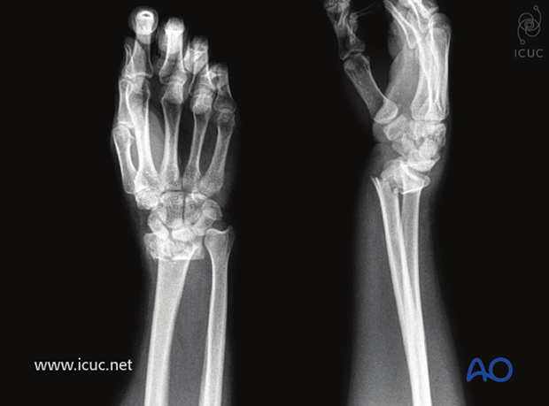
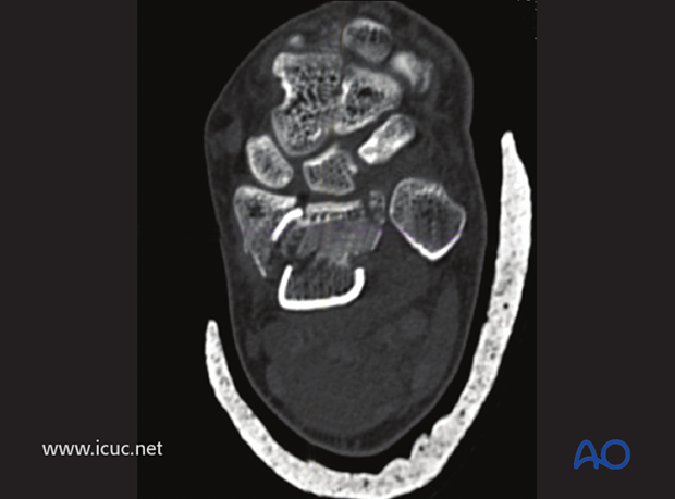
CT images showing fragmentary intraarticular fractures.
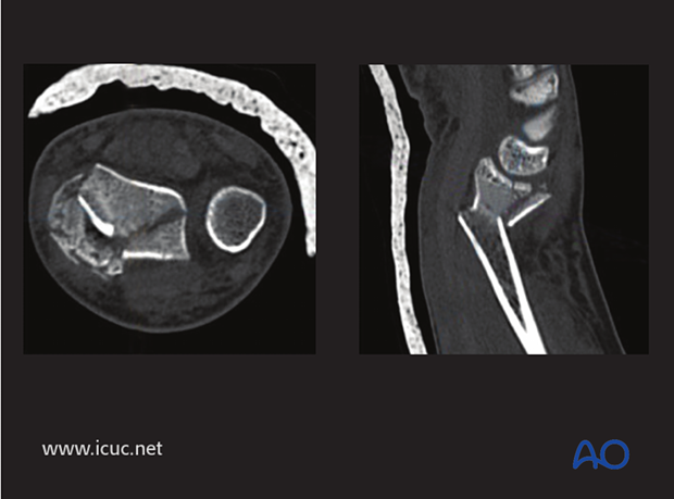
The CT scan always shows more comminution than is shown on a plain X-ray.
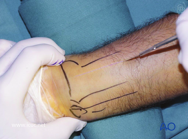
Incision for Henry approach.
Flexor carpal radialis tendon is exposed.
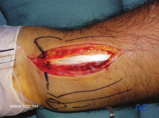
Pronator quadratus is seen beneath the tendon sheath and retracted towards the ulnar side.
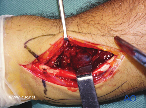
Exposure of the severely displaced distal radial fracture.
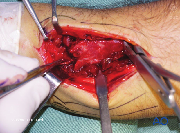
The fracture is cleaned of hematoma and the dorsal fragments are distracted distally to initiate the reduction maneuver.
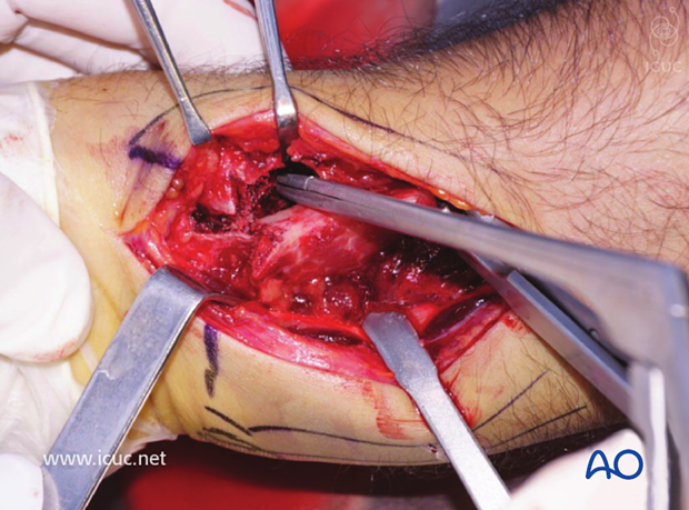
The variable angle plate is positioned once the fracture has been reduced, and confirmed with fluoroscopy.
AP and lateral images showing satisfactory reduction.
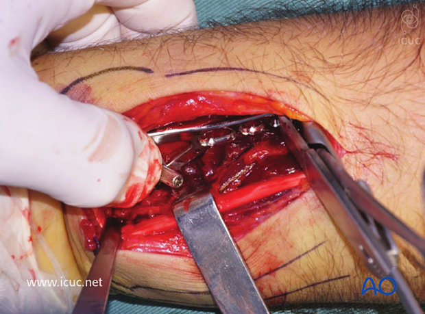
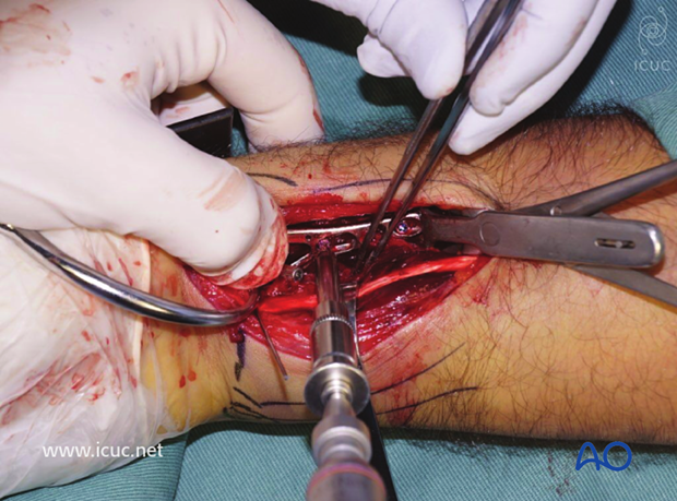
The first screw is inserted into the oblique hole, which allows for subtle plate adjustment proximally or distally.
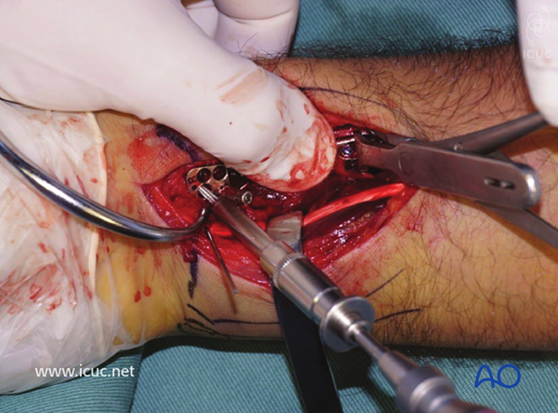
Subchondral screws are inserted in the variable angle plate. Fluoroscopy must be used to ensure they are extraarticular.
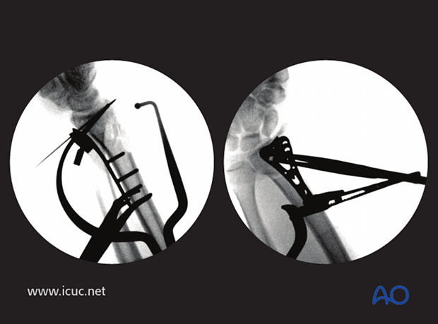
The screws are all extra-articular. Note the dorsal comminution.
Pronator quadratus is closed over the plate.
Final lateral fluoroscopic image
Final AP fluoroscopic image.
Four-week images of healing fracture.
Five-week clinical images. At this stage all movements are limited.
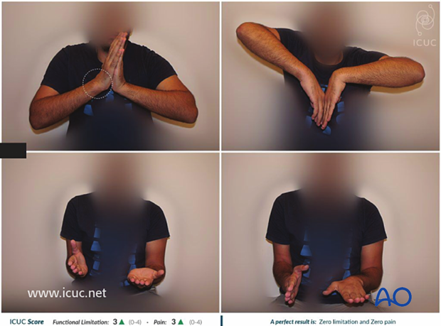
18 week images showing healed fracture.
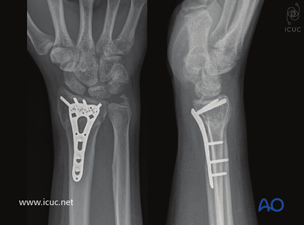
At 18 weeks movement has almost completely returned to normal.
10. Case 2
Displaced, intraarticular distal radial fracture.
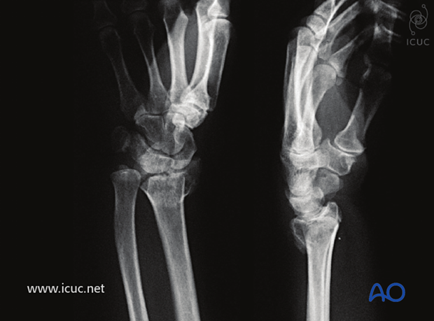
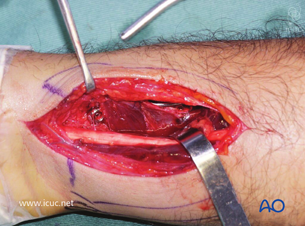
A volar Henry approach was used.
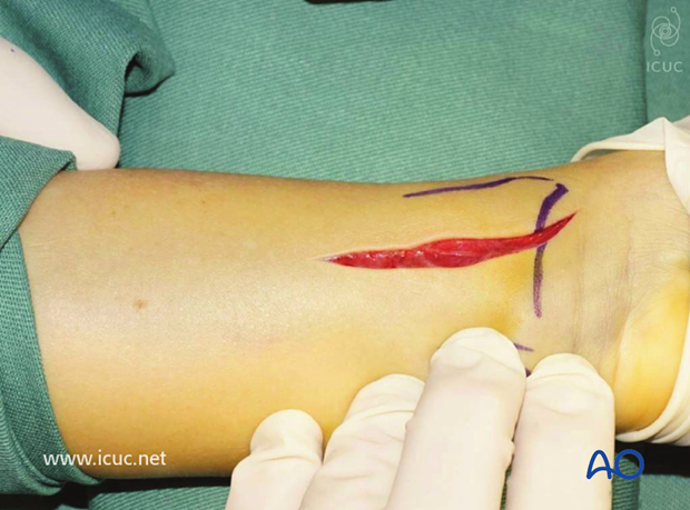
Flexor carpi radialis tendon is retracted towards the ulna and the incision is continued through the volar tendon sheath.
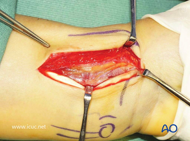
Pronator quadratus has been retracted towards the ulna to show the volar distal radius.
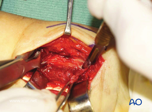
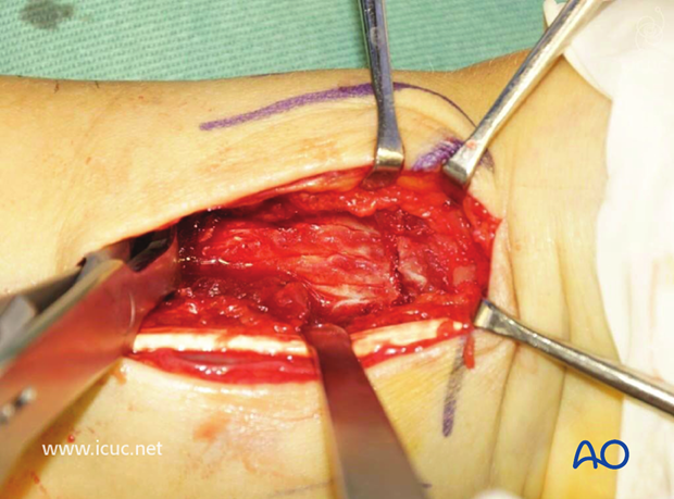
The distal radial fracture is demonstrated and reduced.
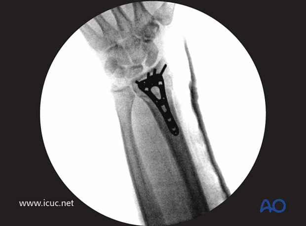
AP fluoroscopic image showing variable angle plate applied in the correct position with K-wire holding plate in place.
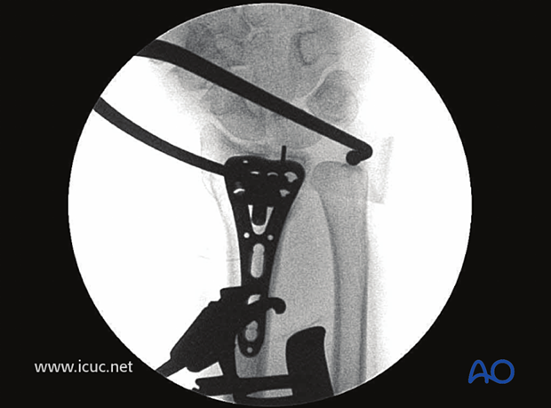
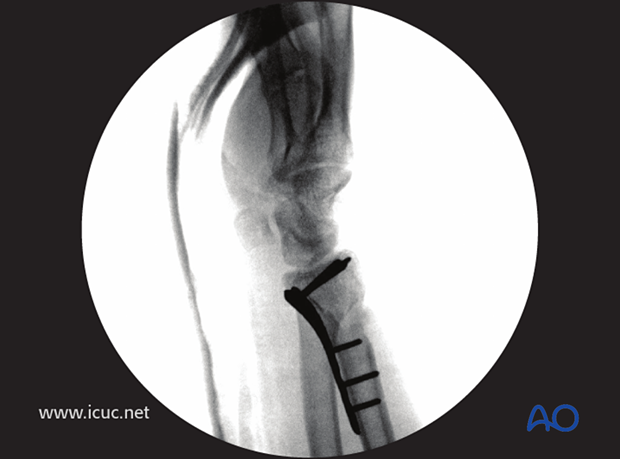
Proximal screws are then inserted.
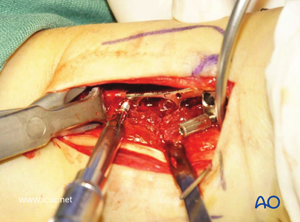
Once reduction has been obtained, the variable angle screws are inserted, under fluoroscopic control to ensure they are extraarticular.
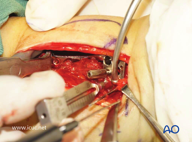
Pronator quadratus is used to cover the plate during closure.
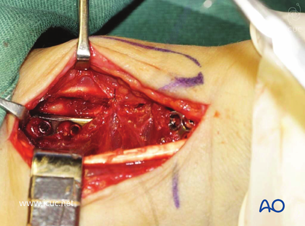
Closed wound.
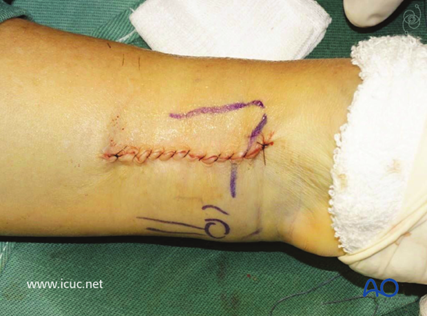
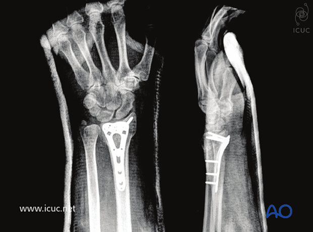
Postopertaive images with the patient in a volar slab showing accurate screw position.
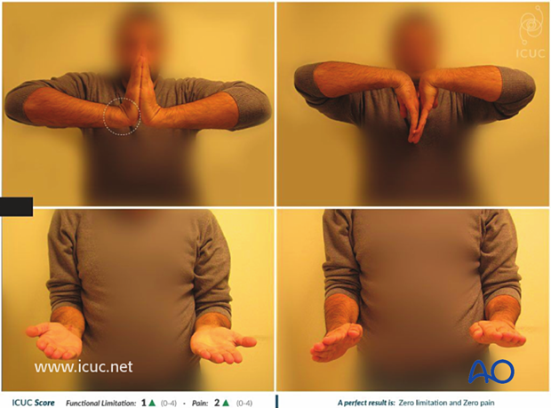
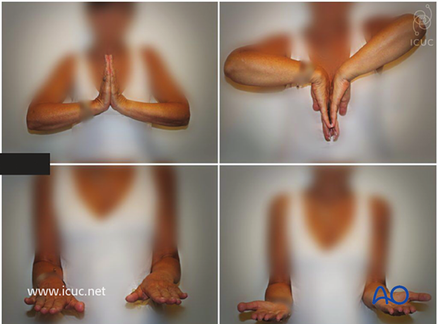
Clinical images showing excellent function at 2 weeks.
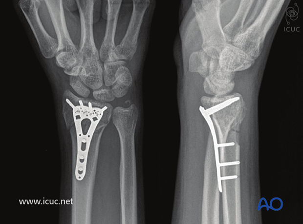
One year images showing healed fracture.
