Palmar approach – modified Henry approach to the distal forearm
1. Palmar approaches
In general, there are two palmar surgical approaches to the distal radius– a modified Henry approach to the radius and a more ulnar approach, designed to expose the median nerve as well as the distal radius.
The modified Henry approach is suitable for most fractures of the distal radius.
If it is desired to decompress the carpal tunnel, this may be performed either through one ulnar extensile approach or two separate approaches.
For isolated lunate facet fractures an ulnar approach is more useful.
For high energy fractures an extended ulnar approach may be used.
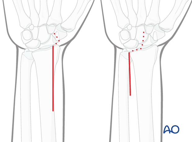
2. Modified Henry approach
The modified Henry approach uses the plane between flexor carpi radialis tendon and the radial artery.
The classical Henry approach goes between brachioradialis and the radial artery, i.e., radial to the radial artery. The modified approach is ulnar to the radial artery.
The flexor carpi radialis tendon is palpated, before making the skin incision to the radial side.
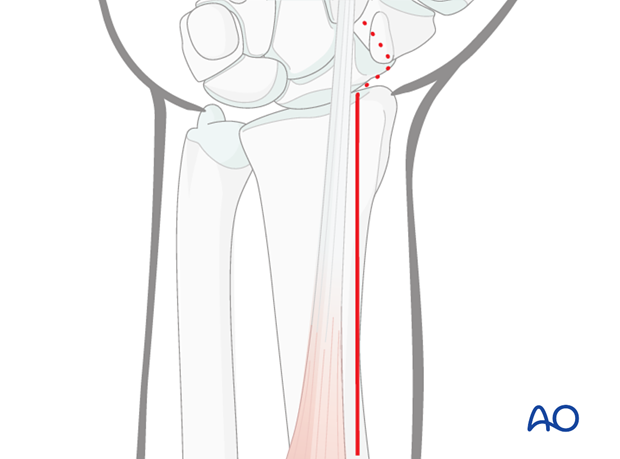
3. Pitfall
The radial artery and the palmar cutaneous branch of the median nerve are at risk during this approach.
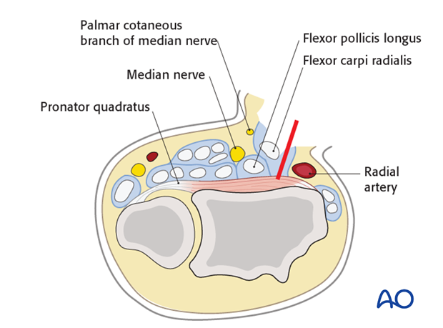
4. Skin incision and exposure
Make the skin incision along the radial border of the flexor carpi radialis tendon.
The sheath is opened and the tendon retracted towards the ulna.
Deepen the incision between the flexor pollicis longus and the radial artery.
Care must be taken to avoid damaging the radial artery on the radial side and the palmar cutaneous branch of the median nerve on the ulnar side.
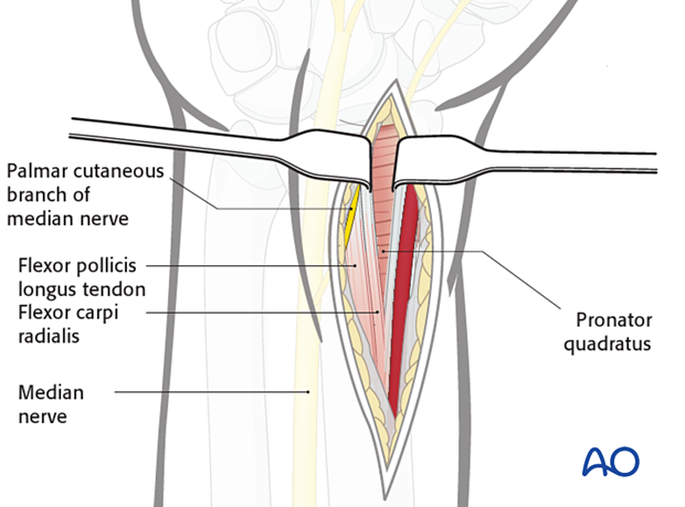
A finger is used to sweep the flexor pollicis longus muscle belly towards the ulna.
This increases the space and exposes the pronator quadratus muscle.
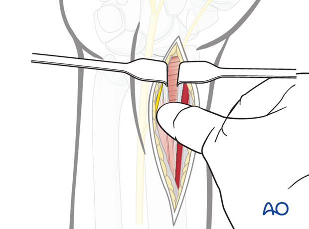
Pearl
The pronator quadratus muscle should be elevated using an L-shaped incision.
The horizontal limb is placed at the watershed line. This lies a few mm proximal to the joint line; the position of the joint line can be determined by a hypodermic needle placed in the joint.
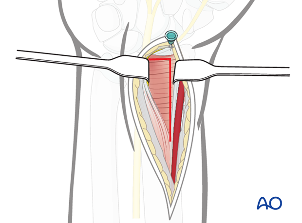
The pronator quadratus muscle is incised on its radial border, exposing the distal radius. It is stripped off the distal radius together with the periosteum.
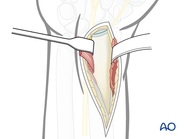
5. Wound Closure
The pronator quadratus should be placed over the plate.
Every attempt should be made to reattach the horizontal limb of the pronator quadratus elevation to the capsule.
If possible, it should be reattached to its radial insertion.
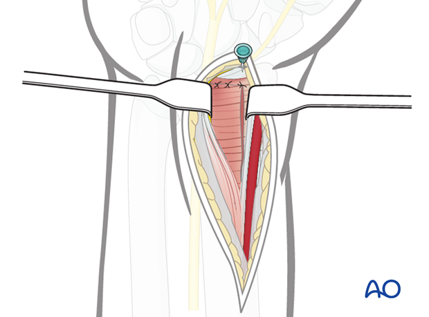
The tendon sheath may be closed, but care must be taken to avoid catching the cutaneous branch of the median nerve.
The skin is closed.


