33B3.2/.3 Partial articular fracture, frontal/coronal, posterior condyle(s)
General considerations
These posterior fractures may be unicondylar or bicondylar.
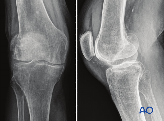
In general, partial articular frontal/coronal fractures are not common. They may be associated with ligamentous injuries to the knee. They are challenging to fix in osteoporotic bone. These fractures are often missed as they are not clearly seen in the AP view. A true lateral view or CT scan is necessary to identify the posterior condylar fragment. Posterior subluxation and knee flexion are most often present during the impact.
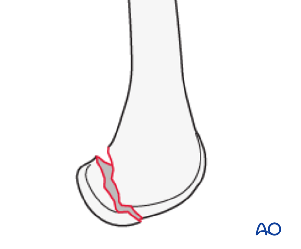
33B3.2 Unicondylar posterior (Hoffa)
This injury may affect either the medial or lateral condyle.

X-ray by courtesy of Spital Davos, Switzerland, Dr C Ryf and Dr A Leumann.

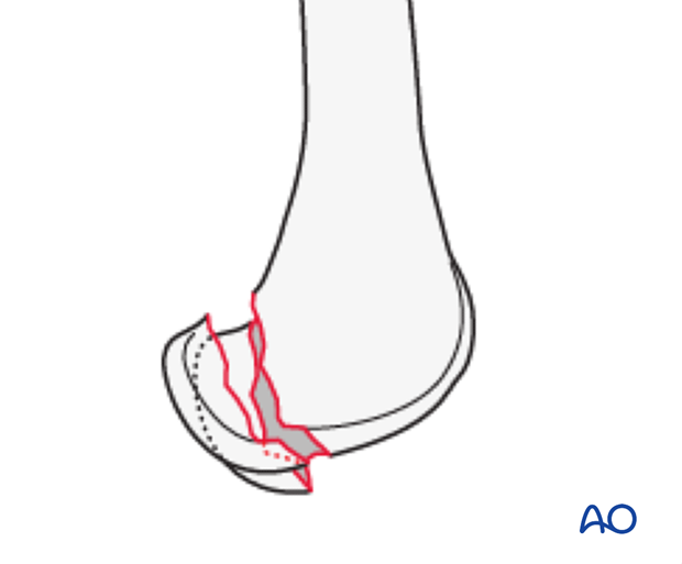
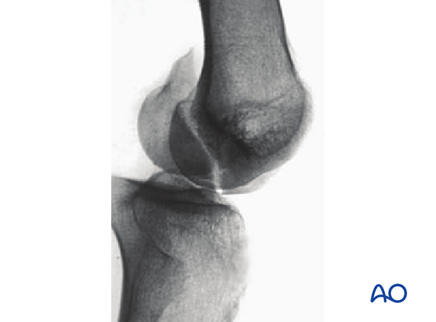
33B3.3 Bicondylar posterior
This injury affect both condyles.

X-ray taken from Orozco R et al, (1998) Atlas of Internal Fixation. Used with kind permission.


