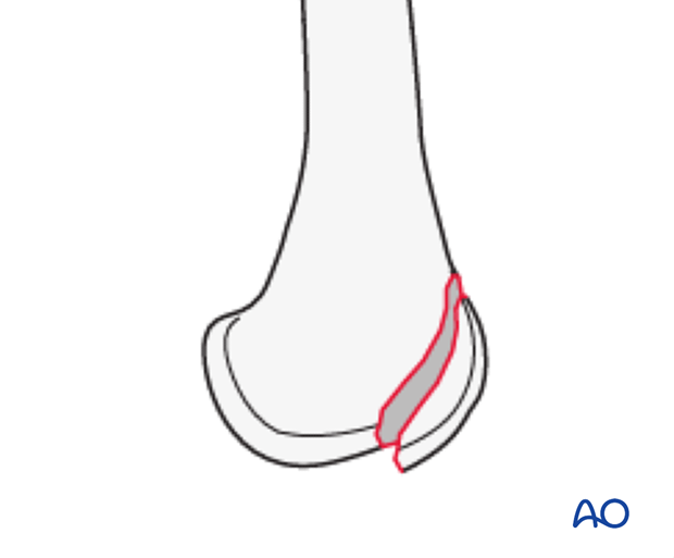
33B3.1 Partial articular fracture, frontal/coronal, anterior and lateral flake
33B3.1 Anterior and lateral flake fracture
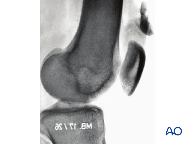
These fractures may be associated with patellar dislocation. These osteochondral fractures may be small and may be technically unfixable. Small fracture fragments may need to be excised, or washed out of the knee joint arthroscopically.
The retropatellar surface must also be checked for osteochondral lesions.
In general, partial articular frontal/coronal fractures are not common. They may be associated with ligamentous injuries to the knee. They are challenging to fix in osteoporotic bone. These fractures are often missed as they are not clearly seen in the AP view. A true lateral view or CT scan is necessary to identify the posterior condylar fragment. Posterior subluxation and knee flexion are most often present during the impact.

X-ray taken from Orozco R et al, (1998) Atlas of Internal Fixation. Used with kind permission.


