Proximal pole fracture of the scaphoid
Definition
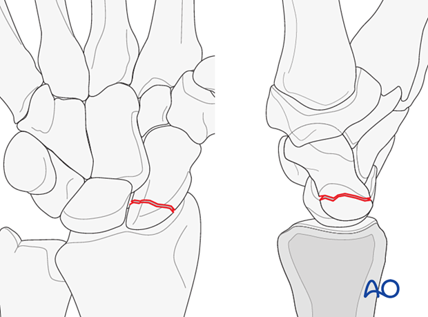
Proximal pole fractures of the scaphoid may be:
- Simple (AO/OTA 72B(a))
- Multifragmentary (AO/OTA 72C(a))
Besides the AO/OTA, the Herbert classification is most often used to describe scaphoid fractures.

Characteristics
Scaphoid fractures are the most common carpal fractures. Debate continues about the management of these fractures. Questions remain about several issues including scaphoid vascularity, clinical and radiological diagnosis, and treatment options.
Imaging
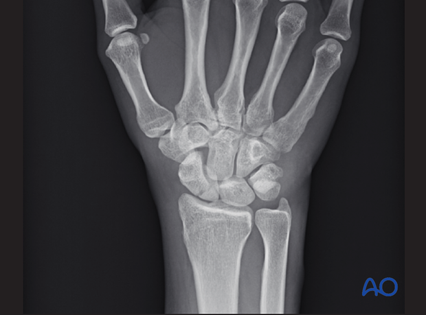
Conventional radiographs do not always adequately demonstrate the fracture configuration.

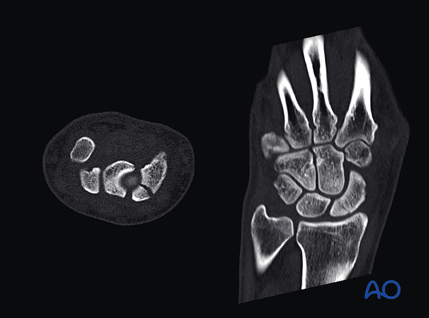
A CT scan is recommended to reveal the degree of displacement.

Pitfall: isolated vs complex carpal injuries
Vascularity
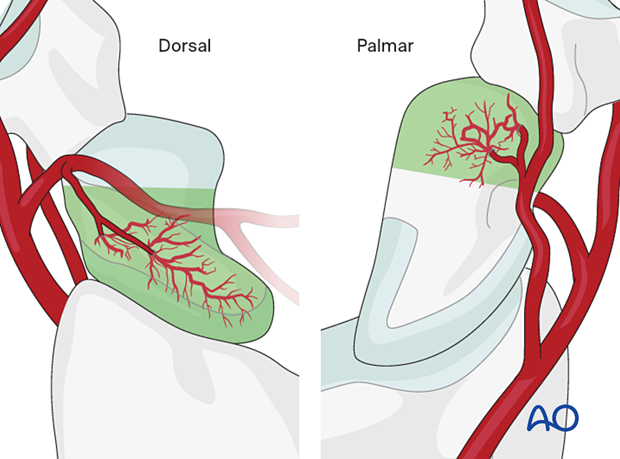
The blood supply of the scaphoid is derived from two sources.
The primary source is a group of vessels entering the dorsal surface of the distal pole. This is the most significant contribution to the vascularity of the scaphoid. For this reason, the proximal pole relies on a retrograde blood flow.
The second group of vessels enters the palmar aspect of the distal pole. These vessels contribute to a lesser degree to the vascularity of the distal third of the scaphoid.
Proximal pole fractures rely mainly on the distal-to-proximal intraosseous blood flow. They are prone to ischemia leading to osteonecrosis, delayed union, and nonunion.


