Greater arc injury/perilunate fracture-dislocation
Definition
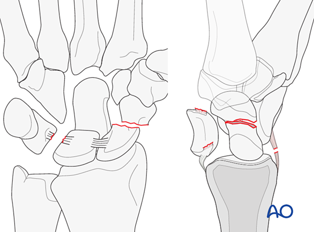
Complex wrist injuries distal to the lunate are usually a combination of fracture and ligamentous injuries. The injury usually starts on the radial side of the carpus and progresses to the ulnar side, depending on the force applied.
Of the greater arc injuries:
- 90% are transscaphoid fracture-dislocations. The triquetrum is usually also fractured
- The second most common injury is transradial styloid fracture-dislocation
There are many bony and ligamentous injury combinations.
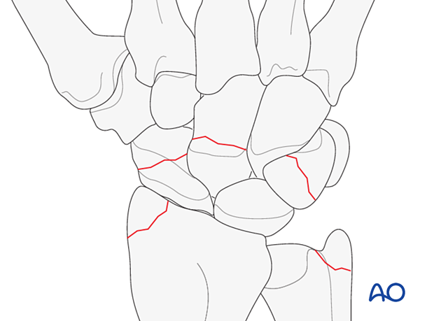
Fractures of the triquetrum, trapezium, trapezoid, and pisiform may be part of complex injuries. These fractures may also appear as isolated injuries. See the entries in the references.

Fractures of adjacent carpal bones may occur, instead of ligamentous ruptures, when the disrupting force propagates around the midcarpal joint.
Concurrent bony and soft-tissue lesions of the carpus are not mutually exclusive (eg, concomitant scaphoid fracture and scapholunate rupture).

Imaging
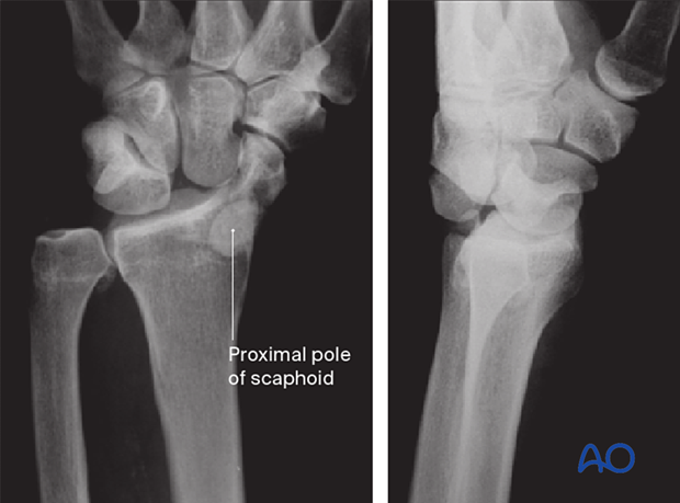
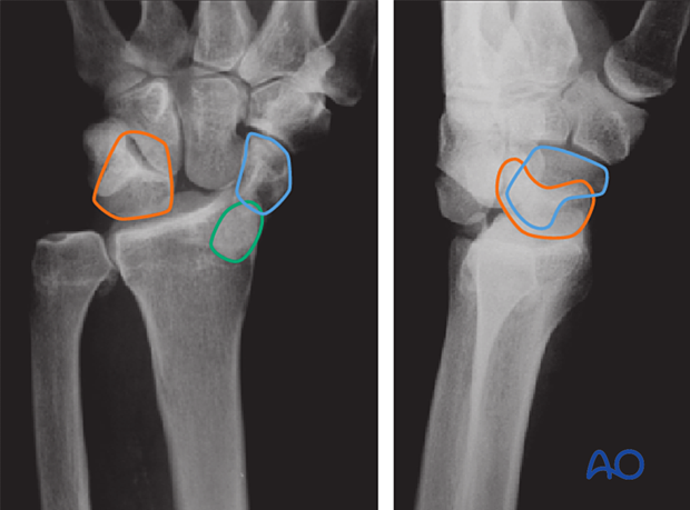
The x-rays of this case show a transscaphoid perilunate fracture-dislocation.

The lines show:
- Lunate (orange)
- Distal scaphoid fragment (blue)
- Proximal scaphoid fragment (green)

Radiological signs in the carpal bones
‘Arcs’ are lines that can be drawn or imagined on x-ray/CT images of the hand and wrist to help assess the alignment of the carpal bones. A discontinuity in an arc indicates a malalignment of the carpal bones either by the fracture or dislocation and should lead to further investigation, eg, CT scan.
Variations of injury patterns can be identified depending on which carpal bones and ligaments are affected and the direction of any dislocation or fracture displacement.
Greater arc injuries comprise fracture-dislocations of the scaphoid, capitate, hamate, and/or triquetrum.
Lesser arc injuries are pure ligamentous injuries around the lunate.
The concept of ‘arcs’ helps to identify the location and extent of a complex carpal injury.


