Lunocapitate and midcarpal dislocation
Definition
Introduction
Lunocapitate dislocation and midcarpal dislocation (stage II and III perilunate injury) are purely ligamentous perilunate dislocations and part of a lesser arc injury.
These injuries result from high-energy trauma.
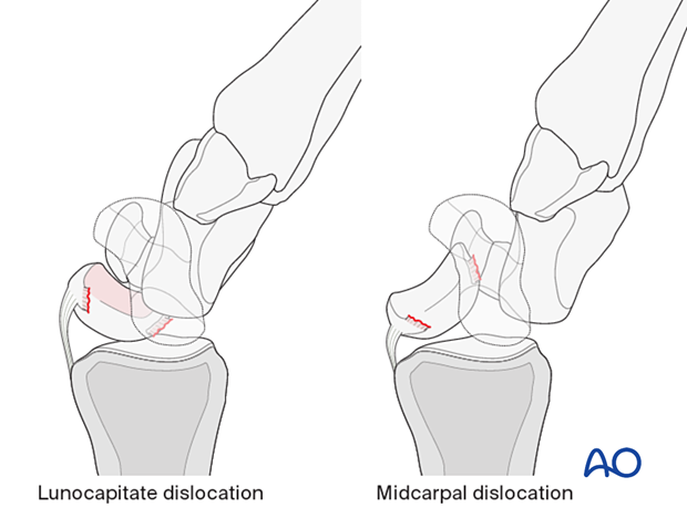
Injury pattern of perilunate dislocations
Pathoanatomy: There is a sequential disruption of carpal ligaments, passing from the radial side to the ulnar side of the wrist. This leads to progressive perilunate instability. The final expression is complete displacement of the carpus from around the lunate.
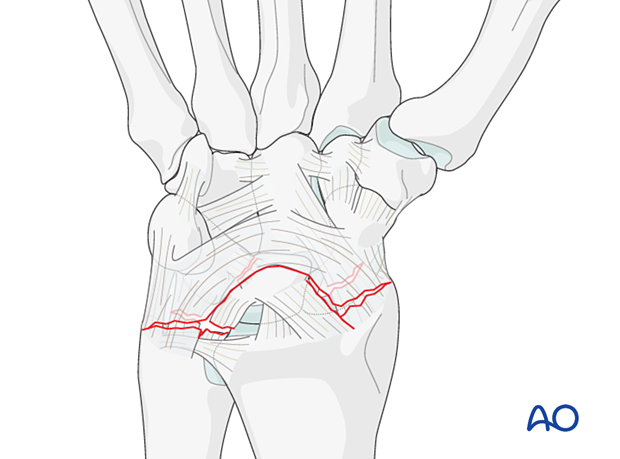
In the lunocapitate dislocation, the lunate stays in line with the forearm, and the rest of the carpal bones are dorsally dislocated.
The scapholunate ligament is disrupted.
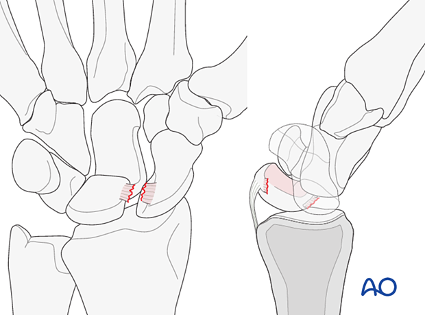
In this more advanced stage of perilunate dislocation, both the scapholunate and lunotriquetral interosseous ligaments fail. A rupture of the palmar extrinsic ligaments also occurs.
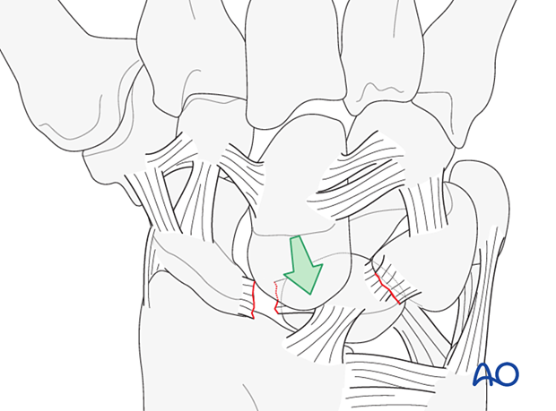
Imaging
This injury may be difficult to diagnose. Initial standard x-rays may not show the extent of injury.
Stress x-rays, CT scans, and MRI scans may be helpful for a complete bone and soft-tissue diagnosis.
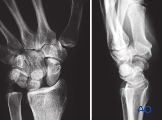
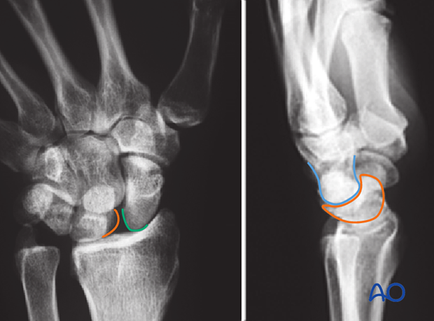
In the AP view, a scapholunate gap is visible. In the lateral view, the tilted lunate orientation indicates a dorsal intercalated segment instability (DISI).
The lines show:
- Proximal scaphoid (green)
- Lunate (orange)
- Capitate (blue)
Radiological signs in the carpal bones
‘Arcs’ are lines that can be drawn or imagined on x-ray/CT images of the hand and wrist to help assess the alignment of the carpal bones. A discontinuity in an arc indicates a malalignment of the carpal bones either by the fracture or dislocation and should lead to further investigation, eg, CT scan.
Variations of injury patterns can be identified depending on which carpal bones and ligaments are affected and the direction of any dislocation or fracture displacement.
Greater arc injuries comprise fracture-dislocations of the scaphoid, capitate, hamate, and/or triquetrum.
Lesser arc injuries are pure ligamentous injuries around the lunate.
The concept of ‘arcs’ helps to identify the location and extent of a complex carpal injury.


