Lunate dislocation
Definition
Introduction
Lunate dislocation (stage IV perilunate injury) is a purely ligamentous injury and part of a lesser arc injury. As the wrist stays in line with the forearm, the lunate is palmarly dislocated.
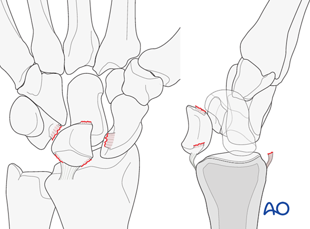
Injury pattern of lunate dislocations
In complete dislocations of the lunate, the displacement is usually in a palmar direction. The greater force required to produce this injury is responsible for massive disruption of both the dorsal and palmar ligaments.
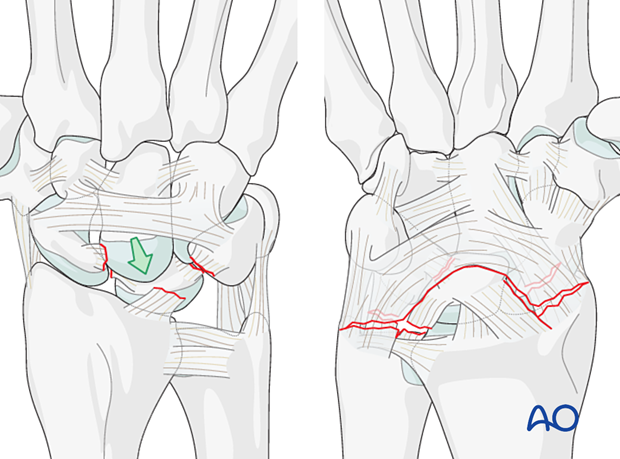
There is also a disruption of the dorsal radiolunotriquetral ligament complex.
Imaging
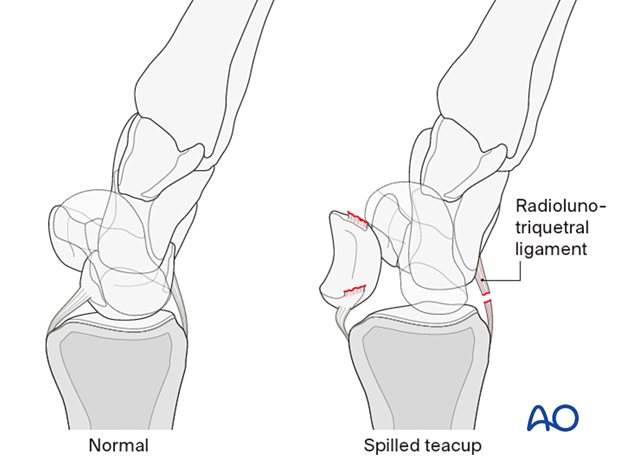
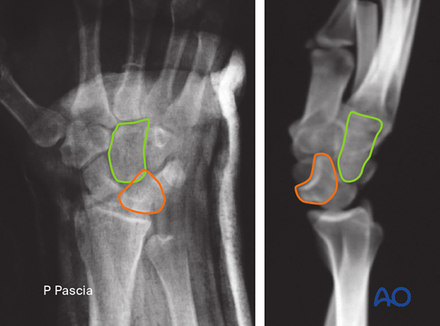
“Spilled teacup” sign
The capitate displaces proximally towards the distal radial articular surface. A lateral x-ray shows the “spilled teacup” configuration of the lunate.
This injury is often missed in an AP x-ray.
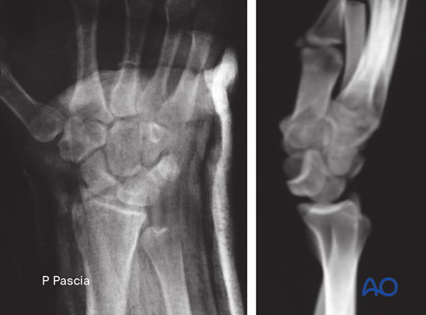
On the AP view, the displaced lunate has a triangular profile rather than its typical quadrilateral image.
The lines show:
- Lunate (orange)
- Capitate (green)
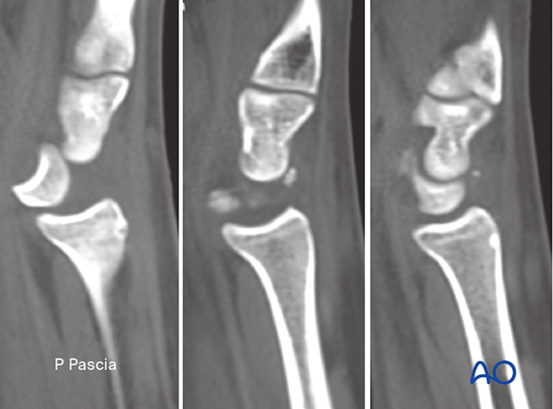
The sagittal 2-D CT scans show the palmar dislocation of the lunate (left) and the empty lunate facet of the radius with some small chip fractures of the lunate (middle). However, there is a normal anatomical relationship between the hamate and the triquetrum (right).
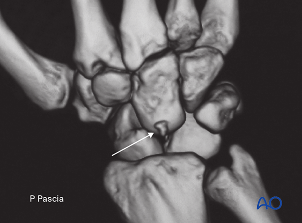
The dorsal view 3-D CT rendering confirms the palmar dislocation of the lunate, although the scaphoid keeps its anatomical relationship with the radius and the distal carpal row. A small chip fracture of the dorsal aspect of the lunate (arrow) suggests an avulsion of the dorsal scapholunate ligament.
Radiological signs in the carpal bones
‘Arcs’ are lines that can be drawn or imagined on x-ray/CT images of the hand and wrist to help assess the alignment of the carpal bones. A discontinuity in an arc indicates a malalignment of the carpal bones either by the fracture or dislocation and should lead to further investigation, eg, CT scan.
Variations of injury patterns can be identified depending on which carpal bones and ligaments are affected and the direction of any dislocation or fracture displacement.
Greater arc injuries comprise fracture-dislocations of the scaphoid, capitate, hamate, and/or triquetrum.
Lesser arc injuries are pure ligamentous injuries around the lunate.
The concept of ‘arcs’ helps to identify the location and extent of a complex carpal injury.


