Ulnar approach to the hamate hook
1. Indication
The ulnar approach to the hamate hook is indicated for excision of the hook. It can also be used for excision of the pisiform in case of posttraumatic sequelae.
In some cases, this approach is used to treat acute fractures.
2. Anatomy
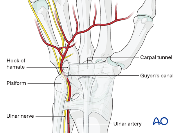
The hook of hamate contributes to the medial border of the carpal tunnel and lateral border of the Guyon’s canal.
The Guyon’s canal is the interval between the hook of the hamate and the pisiform bone and contains the ulnar artery and nerve.
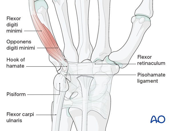
Attachment to the hook:
- Pisohamate ligament
- Flexor retinaculum
- Musculus opponens digiti minimi and flexor digiti minimi
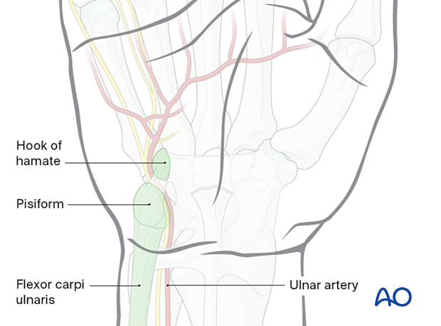
Anatomical landmarks
- Flexor carpi ulnaris tendon on the ulnar side of the wrist, which is inserted into the pisiform bone
- Pisiform bone, palpable distal to the palmar wrist crease
- Ulnar artery palpated radial to the flexor carpi ulnaris (FCU)
- Hook of hamate, palpable distal and radial to the pisiform
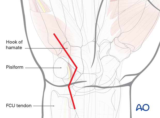
3. Skin incision
Start the zigzag incision at the level of the distal radioulnar joint (DRUJ) radial to the FCU tendon.
It turns radially at the level of the distal palmar crease at the base of the pisiform.
It then turns ulnar-wards and continues distally as shown.
The incision is about 5 cm long in total.

4. Deep dissection
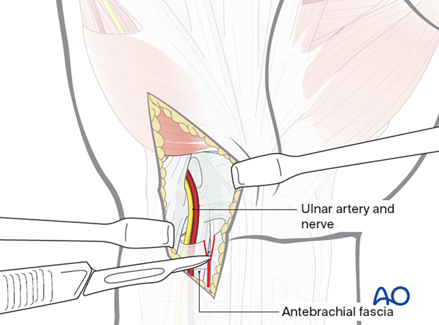
Identify the ulnar artery and nerve in the proximal part of the wound and trace them distally to expose the hook of the hamate.
Divide the fascia antebrachia in the proximal part of the wound.
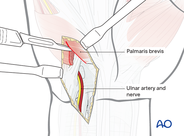
Divide the palmaris brevis distally.
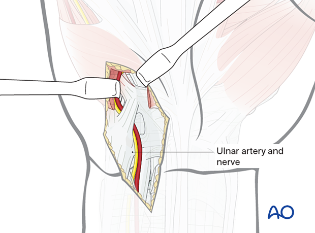
Retract the ulnar artery and nerve to the ulnar side to protect them.
5. Preparation of the hamate hook
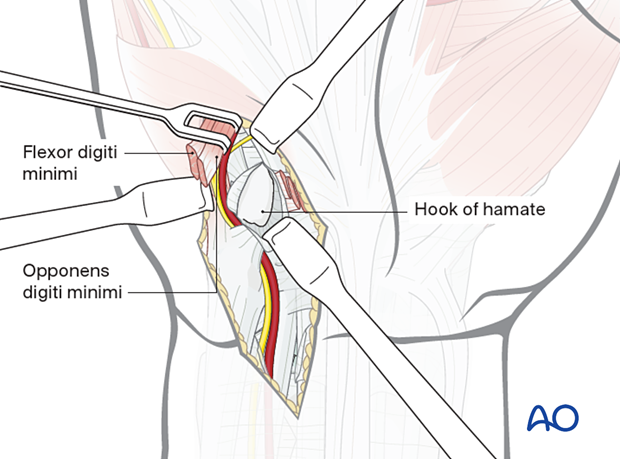
Expose the hook subperiosteally by sharp dissection.
Divide the origin of the flexor digiti minimi brevis (and opponens digiti minimi) and retract it to the ulnar side.
On the radial side of the wound, the flexor digiti minimi tendon can be identified.
6. Wound closure
Close the skin in a standard manner.


