Palmar approach to the scaphoid
1. Indications
The palmar approach to the scaphoid is indicated for the following injuries:
- Irreducible, displaced scaphoid fractures in the distal two thirds
- Comminuted scaphoid fractures
AO teaching video
Wrist joint–Scaphoid bone – Palmar approach
2. Skin incision
Anatomical landmarks
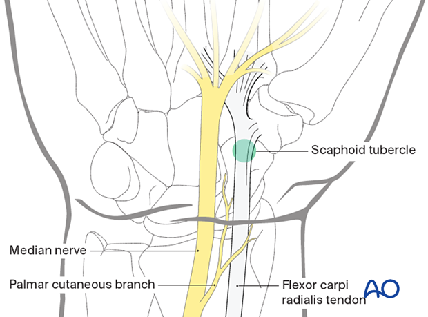
The landmarks for this incision are:
- Scaphoid tubercle
- Flexor carpi radialis (FCR) tendon
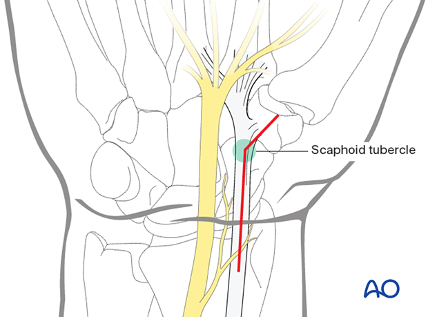
Angled skin incision
The incision is in line over the distal part of the FCR tendon and then turns at the level of the palmar rim of the distal radius towards the scaphoid tubercle and scaphotrapezial joint.

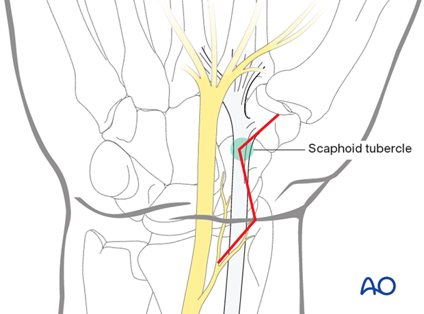
Zigzag incision
Alternatively, a zigzag incision may be constructed using the same landmarks. This incision may result in better cosmetic outcome in case of very pronounced palmar crease.
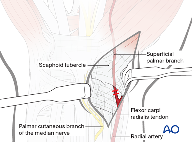
3. Ligation of the superficial palmar branch of the radial artery
The superficial palmar branch of the radial artery passes towards the palm, running close to the scaphoid tubercle. If necessary, it can be ligated and divided.
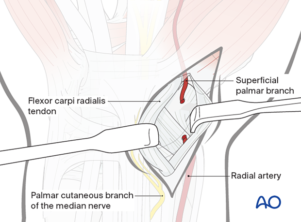
4. Opening the FCR sheath
Open the FCR sheath as far distally as possible and retract the tendon towards the ulnar side.
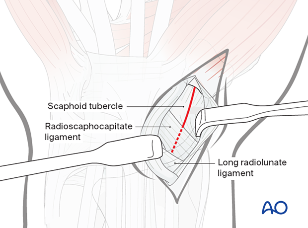
5. Exposure of the wrist capsule
Open the capsule obliquely, starting at the scaphoid tubercle towards the palmar rim of the radius.
As determined by the fracture configuration, preserve as much of the palmar ligament complex as possible. This helps to contain the proximal pole and prevent palmar tilt of the scaphoid.
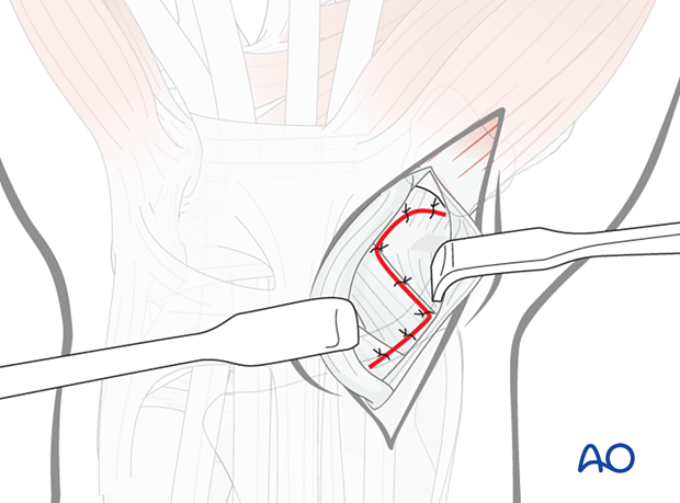
Alternatively, a zigzag incision may be used. It starts at the level of the scaphoid tubercle distally and turns in line with the radioscaphocapitate ligament. It then turns at the level of the radial styloid down to the pronator quadratus.
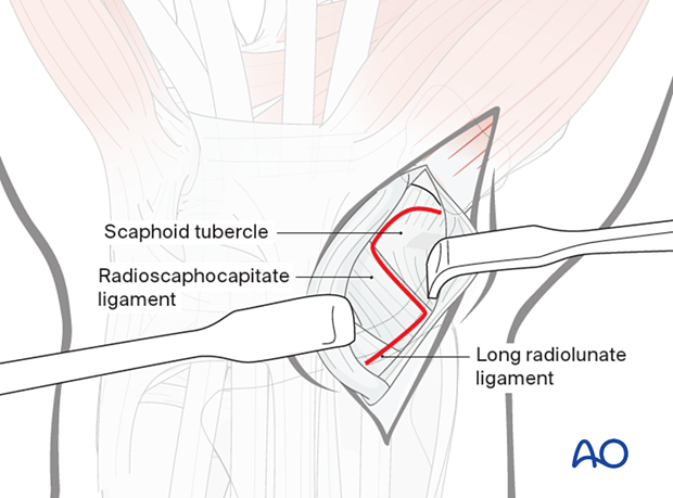
6. Exposure of the scaphoid
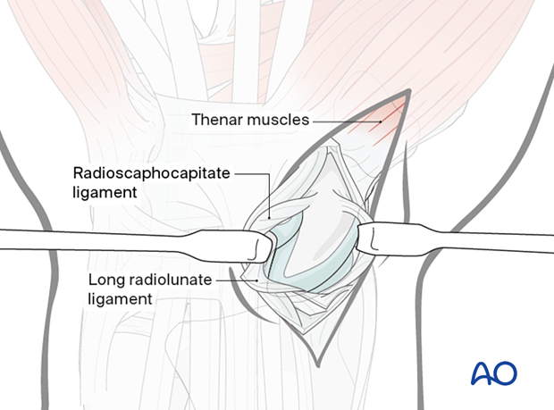
Retract the divided radioscaphocapitate ligament to expose the scaphoid.
If it is necessary to expose the proximal part of the scaphoid, divide the long radiolunate ligament proximally as far as the palmar rim of the radius.
7. Exposure of scaphotrapezial joint
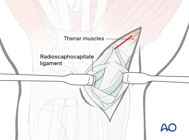
Expose the scaphotrapezial joint to allow optimal positioning of a screw.
Deepen the incision distally, dividing the origin of the thenar muscles in line with their fibers.
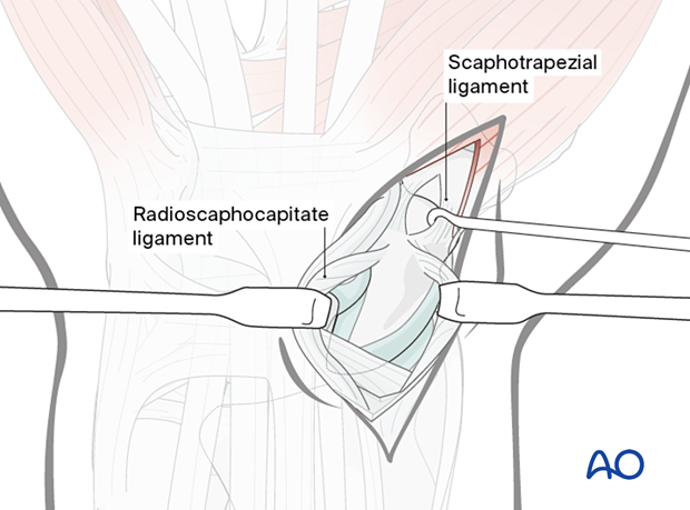
Identify the scaphotrapezial joint, divide the scaphotrapezial ligament in the line of its fibers, and open the joint capsule.
8. Wound closure
To prevent secondary carpal instability, the divided palmar ligaments (radioscaphocapitate/long radiolunate) must be repaired with fine interrupted sutures.
Approximate the soft tissues over the scaphotrapezial joint.
Test the integrity of the soft-tissue repair by passive wrist motion.
Finally, the FCR tendon sheath is repaired and covered with subcutaneous tissue.
