Minimally invasive palmar approach to the scaphoid
1. Indications for this approach
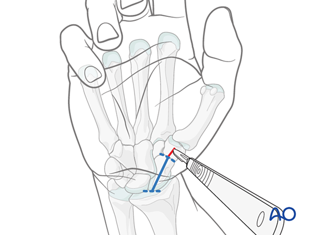
A minimally invasive palmar approach to the scaphoid is used for screw fixation of minimally displaced fractures of the scaphoid waist (<2 mm).
In minimally displaced scaphoid fractures of the proximal third (<1 mm), this may also be an option.
AO teaching video
Scaphoid-Fracture – Percutaneous Fixation with the 3.0 mm Headless Compression Screw (HCS)
At 9:28, a clinical case shows the steps for percutaneous screw fixation of a scaphoid fracture.
2. Intraoperative imaging
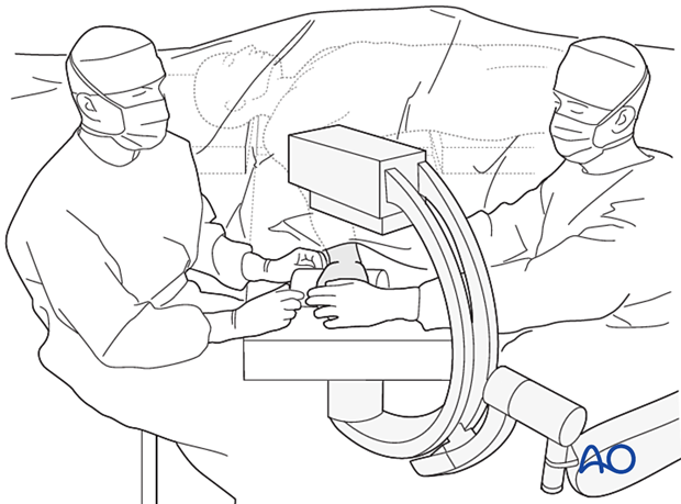
When accessing the scaphoid percutaneously, the use of an image intensifier is essential.
3. Confirming the fracture pattern
Before starting a percutaneous procedure, screen the scaphoid with an image intensifier to confirm that the fracture is suitable for this type of fixation.
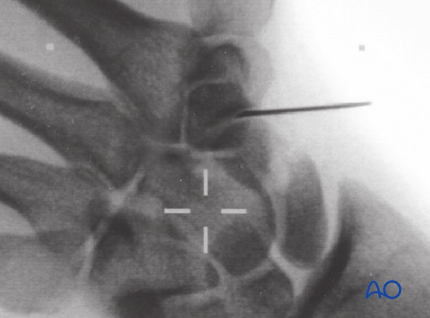
It can be helpful to mark the scaphotrapezial joint space with a hypodermic needle or fine K-wire placed percutaneously under the image intensifier.
4. Hyperextension
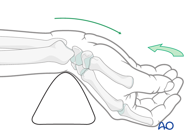
Place a rolled towel or bolster under the wrist and hyperextend it.
This maneuver helps to:
- Indirectly reduce the scaphoid fracture by ligamentotaxis
- Present the distal pole of the scaphoid more readily when the scaphotrapezial joint is opened
5. Marking the skin
It may be helpful to mark on the skin the position of the scaphoid, the palmar rim of the distal radius, and the level of the scaphotrapezial joint.
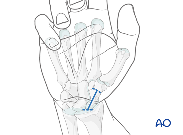
6. Skin incision
A stab incision of about 1 cm long is made distal to the scaphotrapezial joint.
The subcutaneous tissues are dissected down to the capsule of the scaphotrapezial joint. The capsule is incised.
The distal pole of the scaphoid is now accessible for insertion of a guide wire.


