Dorsal approach to the scaphoid
1. Indications
The dorsal approach to the scaphoid is used for the following injuries:
- Proximal pole fractures
- Complete scapholunate (SL) ligament rupture
- Scaphoid fractures or complete SL ligament ruptures with concomitant distal radial fractures
2. Surgical anatomy
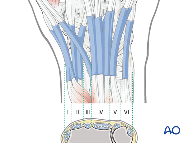
There are six extensor compartments on the dorsum of the radiocarpal region:
- The 1st compartment contains the tendons of abductor pollicis longus (APL) and extensor pollicis brevis (EPB)
- The 2nd compartment contains the extensors carpi radialis longus and brevis (ECRL and ECRB, respectively)
- The 3rd compartment contains the tendon of the extensor pollicis longus (EPL)
- The 4th compartment contains the tendons of the extensor digitorum
- The 5th compartment contains the tendon of the extensor digiti minimi
- The 6th compartment contains the tendon of the extensor carpi ulnaris (ECU)
3. Skin incision
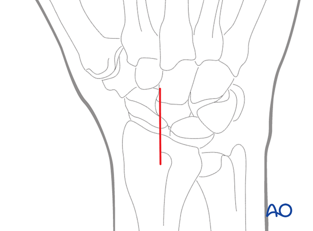
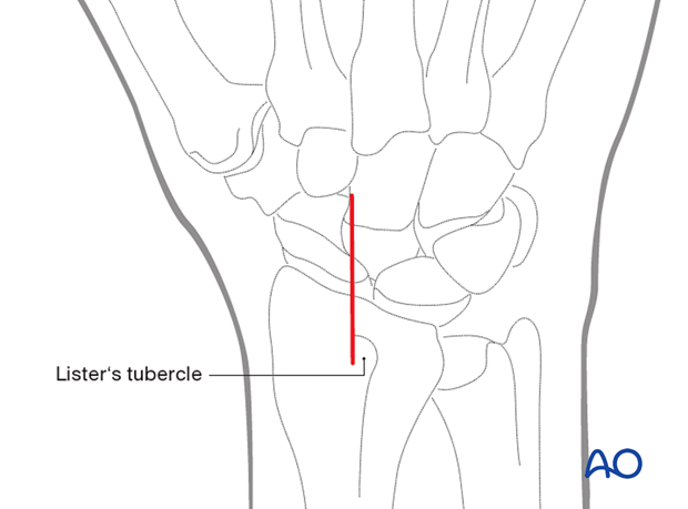
Make a straight dorsal skin incision starting over Lister’s tubercle and extending for about 3 cm distally.
4. Identifying the radial nerve
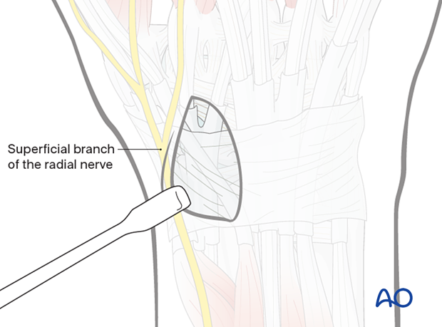
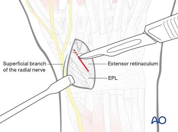
Identify and preserve the superficial dorsal branch of the radial nerve, which runs in the radial skin flap of the wound.
5. Incision of the retinaculum
Incise the extensor retinaculum over the extensor pollicis longus (EPL) tendon, opening the distal part of the 3rd extensor compartment.
6. Retraction of the tendons
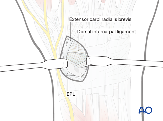
The EPL tendon is then retracted radially together with the tendons of the 2nd extensor compartment (extensor carpi radialis brevis and longus).
The 4th extensor compartment, containing the extensor digitorum and extensor indicis, is located on the ulnar side.
7. Opening the capsule
Make a longitudinal or T-shaped incision, starting at the dorsal rim of the distal radius, and extending to the dorsal intercarpal ligament.
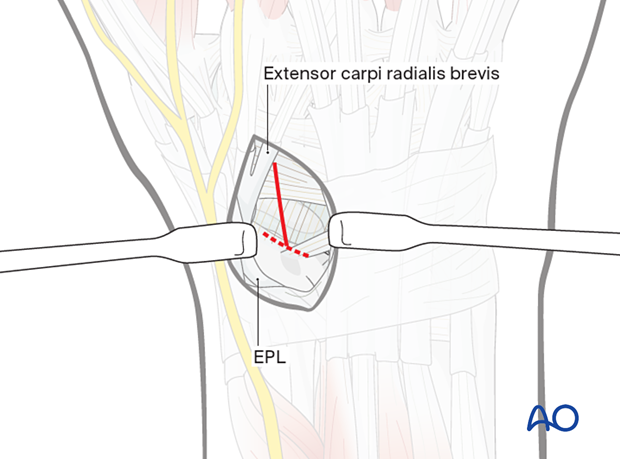
Take care to preserve the vessels to the dorsal ridge of the scaphoid.
The capsule is not stripped from this area.
8. Exposure of the scaphoid
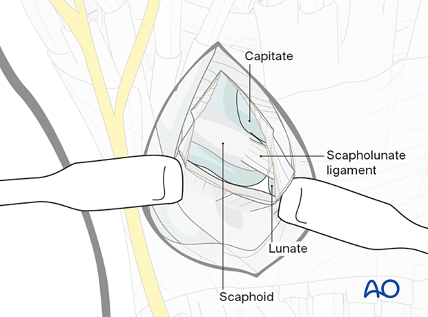
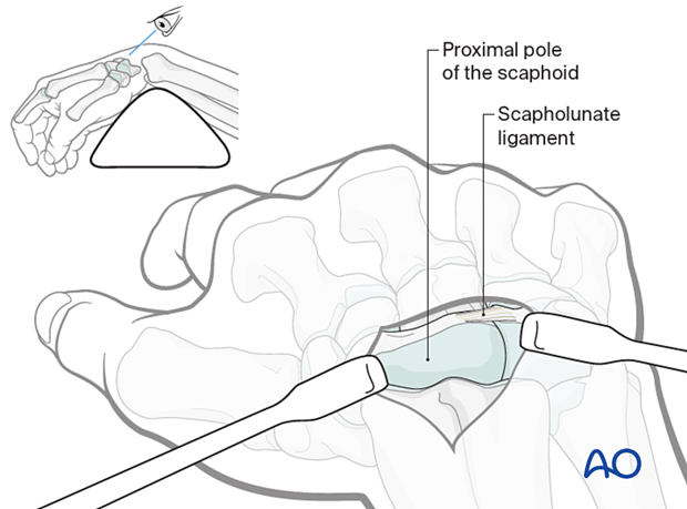
To expose the proximal pole of the scaphoid, it is necessary to flex the wrist.
The scaphoid now comes into view. Identify the SL ligament.
9. Wound closure
Close the capsule with interrupted sutures.
Close the 3rd extensor compartment, avoiding any tension over the EPL tendon, which must glide smoothly. If this is not possible, the EPL tendon is best left superficial to the retinaculum in the subcutaneous tissue.


