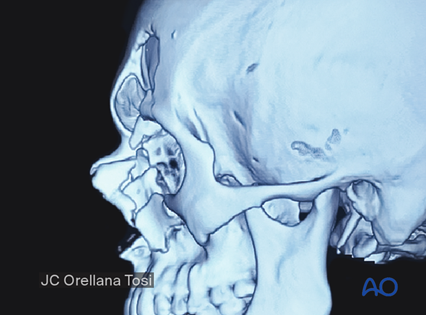
NOE Type III fracture
Definition
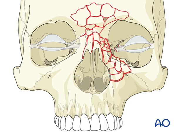
There is often comminution of the NOE area in type III fractures (as in type II fractures). In Type III fractures, the medial canthal tendon is avulsed from its bony insertion.
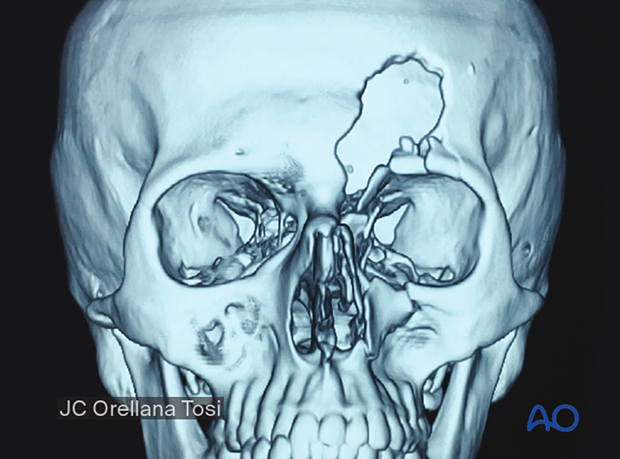
The image shows a unilateral NOE type III fracture.
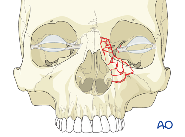
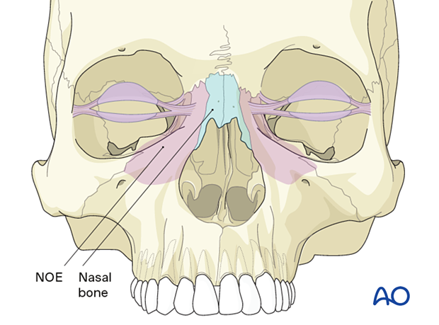
Involvement of the nasal bone
The nasal bones are usually involved and may not provide adequate dorsal support to the nasal bridge. In such cases, in addition to piecing the nasal bone fracture together with wires or mini-screws, bone graft reconstruction is often indicated.
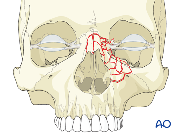
Bilateral type III fracture with nasal bone involvement
The illustration shows a bilateral NOE type III fracture. The nasal bones and cartilages are usually involved and may be severely injured. Bone grafting of the nasal dorsum and caudal nose is typically necessary.
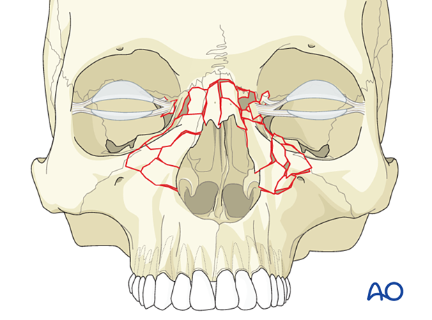
Association with frontal sinus fractures
NOE fractures are often associated with frontal sinus and other upper facial fractures such as fractures of the zygoma and maxilla.
General
The nasoorbitoethmoid (NOE) fracture refers to injuries involving the area of confluence of the nose, orbit, ethmoids, the base of the frontal sinus, and the floor of the anterior cranial base. The area includes the insertion of the medial canthal tendon(s). NOE fractures, by definition, are a different entity to isolated nasal bone fractures. However, they are often associated with fractures of the nasal bones.
NOE fractures are most commonly classified as:
- Type I
- Type II
- Type III
These can be unilateral or bilateral injuries.
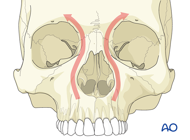
NOE complex fractures involve the medial vertical (nasomaxillary) buttresses of the facial skeleton. Click here for further details on buttresses.
Radiographic findings
Gunshot wound
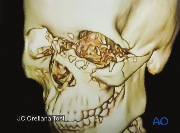
This 3D CT scan is a lateral view of a gunshot wound case with a comminuted fracture. The medial canthal ligament is presumably detached from its bony attachment.
Association with frontal sinus fractures
This 3D CT scan shows a patient with a comminuted fracture. The medial canthal ligament may be detached from a significant bone fragment.
Note the association with the frontal sinus fracture.
Lateral view of the same patient.
