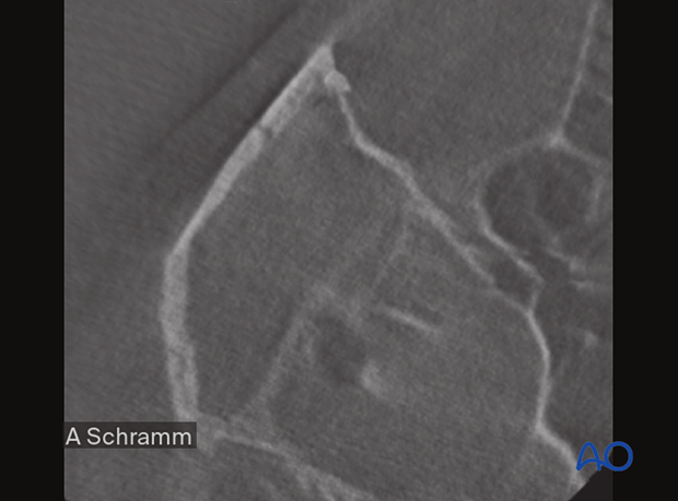
CAS: intraoperative imaging in closed treatment
1. Introduction
Indications
Whenever an intraoperative CT scanner is available, an intraoperative scan should be obtained for intraoperative evaluation of the reduction.
An intraoperative CT scan has the following advantages:
- Avoids surgical revisions
- Eliminates cost of the second procedure
An intraoperative CT scan has the following disadvantage:
- Increased costs
When using computer assisted surgery (CAS), reduction (and fixation if necessary) is performed according to standard procedures described in the AO Surgery Reference. CAS should be considered an adjunct to surgical treatment.
When treating fractures CAS allows intraoperative visualization of the reduction using intraoperative imaging combined with image fusion of preoperative and intraoperative CT scans.
With this technique, insufficient fracture reduction can be identified and corrected, eliminating the need for a return to the OR that may be necessary if only postoperative imaging is performed.
Intraoperative imaging requires an additional 10–15 minutes.
2. Preoperative imaging
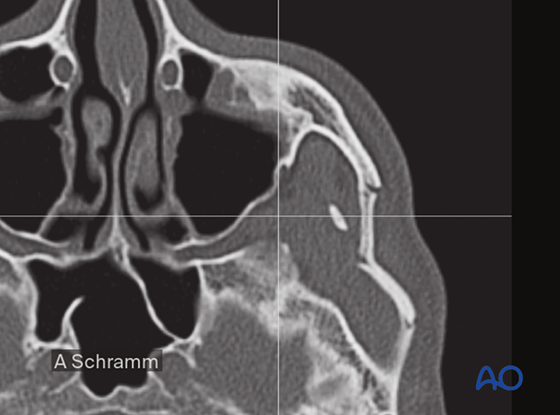
A CT scan from vertex to menton with 1 mm slices is the gold standard for the evaluation of zygomatic bone/midface fractures. Isolated zygomatic arch fractures are not very common, so the entire face should be scanned for the presence of facial trauma. This will help the surgeon avoid missing fractures at other zygomatic bone processes and the associated orbital floor.
The preoperative CT scan shows a displaced isolated zygomatic arch fracture of the left side.
3. Intraoperative assessment of reduction
CT scans can be performed intraoperatively to verify that the zygomatic arch has been properly reduced.
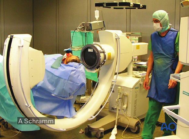
The intraoperative CT scan shows anatomic reduction of the bony fragments, which appear to be stable.
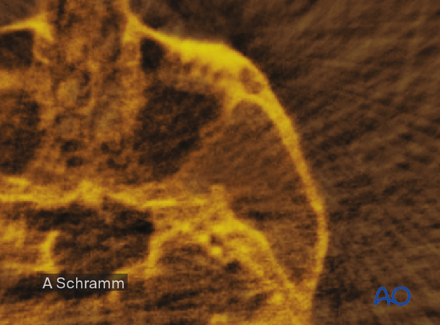
Virtual planning software allows automatic image fusion of preoperative (blue) and intraoperative (gold) CT scans. This allows for better visualization of the achieved reduction.
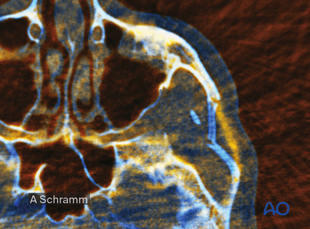
In the example shown here, the intraoperative imaging shows an incomplete reduction of the right zygomatic arch.
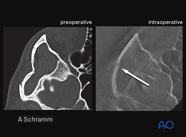
Without any additional morbidity, the reduction of the zygomatic arch could be completed during the same operative session and verified with a second intraoperative scan.
