Transoral approach to the angle
1. Principles
Vestibular incisions
The transoral approach is used for the majority of simple angle fractures.
There are two surgical approaches depending on whether or not a third molar is to be extracted.
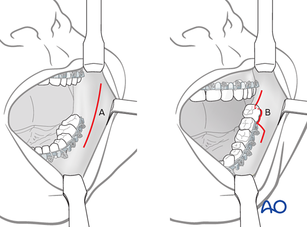
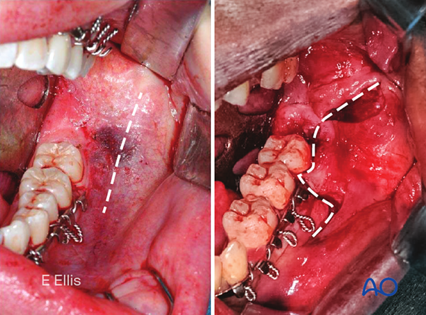
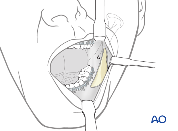
When there is no third molar present, or where one is present but is to be left in place, a purely vestibular incision approximately 5 mm away from the attached gingiva is made (A).
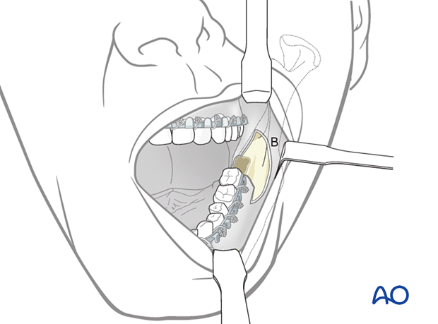
When an erupted the third molar is to be removed, the incision must incorporate a sulcular incision around the tooth's buccal side (B, the combination of vestibular and envelope incisions).
Oral contamination is not a contraindication for an intraoral incision.
Restricted access and contamination
In complex fractures, including comminuted, edentulous, and avulsive fractures that will require the placement of load-bearing reconstruction plates, a transfacial/transcutaneous approach can provide better access to treat the injury and place the internal fixation hardware.
2. Anatomical considerations
Sensory buccal nerve
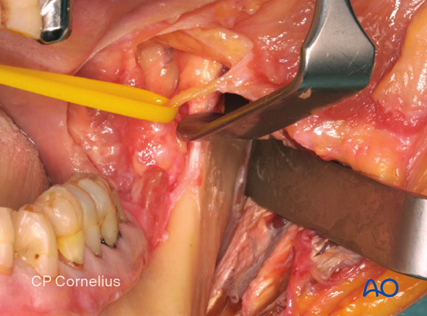
The buccal sensory nerve crosses the upper anterior rim of the mandibular ascending ramus in the coronoid notch region. It is usually below the mucosa running above the temporalis muscle fibers. When the posterior vestibular incision is carried sharply along the bony rim, the buccal nerve is at risk of transection, followed by numbness in the buccal mucosal region. Therefore, to protect the nerve, the posterior dissection is extended bluntly as soon as the lower coronoid notch is reached.
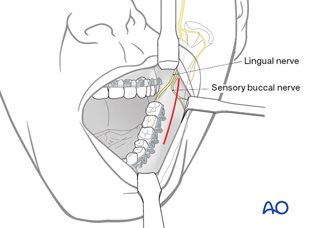
The photograph shows the buccal sensory nerve.
Buccinator muscle
The lateral mucogingival vestibular incision transects the lower attachment of the buccinator muscle. Lateral stripping the mucoperiosteal flap disrupts the attachment of the muscle. To reattach the muscle, the sutures for wound closure in the lateral vestibular should not only be superficial. The suture should catch all layers (mucosa and muscle) as a safeguard for muscle reattachment.
The buccinator muscle belongs to the mimetic muscle system and has a unique functional structure allowing for a movement comparable to a peristaltic motion. The deep fibers run in parallel bundles from the modiolus to the pterygomandibular raphe at the occlusal plane's level. They account for the buccinator mechanism building up a ridge towards the occlusal plane. Its detachment can result in an impaired bolus transport out of the buccal space, which is troublesome for the patient. The buccinator is innervated by the buccal motor branch of the facial nerve.

3. Vestibular incision
Unless contraindicated, infiltrate the area with a local anesthetic containing a vasoconstrictor.
Variant A
Make an incision through the mucosa in the vestibule approximately 5 mm away from the attached gingiva (in the mucogingival junction), extending up the external oblique ridge.
Variant B
Make an incision through the mucosa in the vestibule to incorporate a sulcular incision around the tooth's buccal side of the third molar (the combination of vestibular and envelope incisions).
(Here the third molar has already been extracted to better illustrate the incision design.)
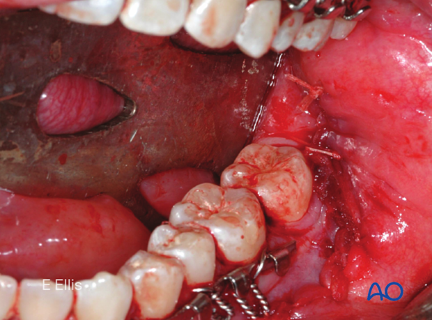
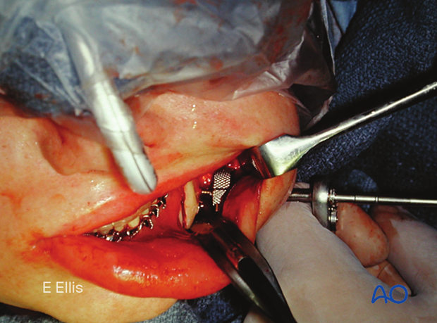
This clinical image shows the fracture exposed, reduced, and MMF secured. Note that the erupted third molar has been removed, and the surgical approach shown here is variant B.

4. Combination with the transbuccal technique
The transbuccal trocar may also assist the surgeon in positioning posterior and inferior screws, sometimes avoiding the need for a transcutaneous approach.
- A detailed description of the transbuccal technique.
5. Wound closure of the vestibular incision
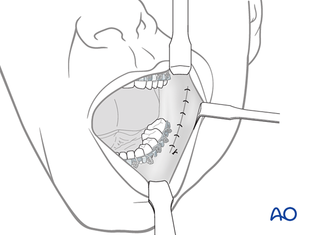
Closure of variant A
After thoroughly irrigating the wound and checking for hemostasis, the incision is closed using interrupted or running resorbable sutures.
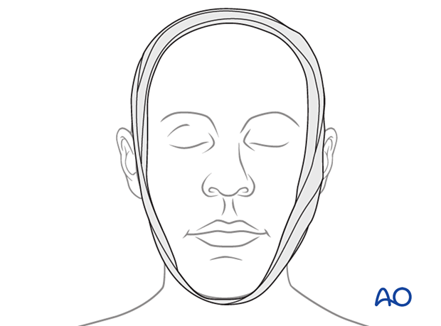
An elastic pressure dressing covering the angle region helps support the soft tissues and prevent hematoma formation.
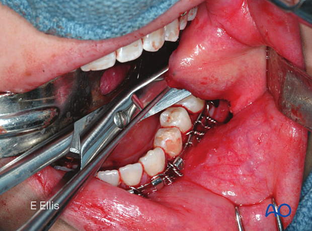
Closure variant B
The cheek flap is undermined with scissors to facilitate tension-free advancement over the extraction site. Generally, resorbable sutures are used for this closure.
The periosteum can be scored or incised to release the flap and allow for tension-free closure across the extraction site.
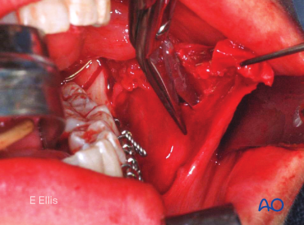
The flap is advanced and closed over the extraction site.
