Upper eyelid weight
1. Introduction
Currently there are no good solutions for dynamic reconstruction of the upper eyelid. Techniques involving springs, magnets, local muscle flaps, and free flaps have all failed to provide predictable eyelid closure.
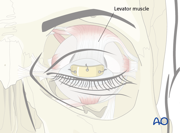
The most reliable closure technique has involved placement of a weight within the upper eyelid, allowing closure to occur by gravity. With this technique, the patient learns to relax the levator muscle, allowing the weight to bring the upper eyelid down.
2. Planning and surgical preparation
Implant selection
The selection of the weight size is a balance between adequate closure to protect the cornea reducing exposure symptoms and upper eyelid ptosis.
The two common materials for weights are gold and platinum. Platinum has a smaller size and thinner profile for less visibility.

Surgical preparation
Clinical examination is the best way to choose the appropriate eyelid weight.
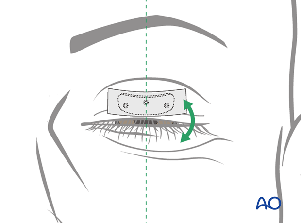
Different weights are placed on the skin centered over the mid-pupillary line of the patient's upper eyelid and taped in position.
The patient is encouraged to open and close the eyelid.
The amount of closure compared to ptosis is judged and the best weight is selected.
When considering the optimal weight, there is a tradeoff between functional closure and eyelid position and appearance during opening.
If considering lower lid suspension, then upper lid procedures should be considered secondarily. Lower lid procedures can alter the size of upper eyelid weight required.
Surgical site marking
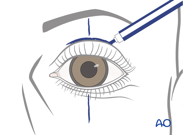
The mid pupillary line as well as the supratarsal fold is marked out with the patient in a sitting position prior to the procedure.
3. Technique
This procedure can be performed either under local or general anesthetic.
Approach
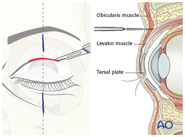
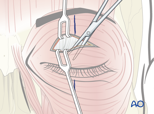
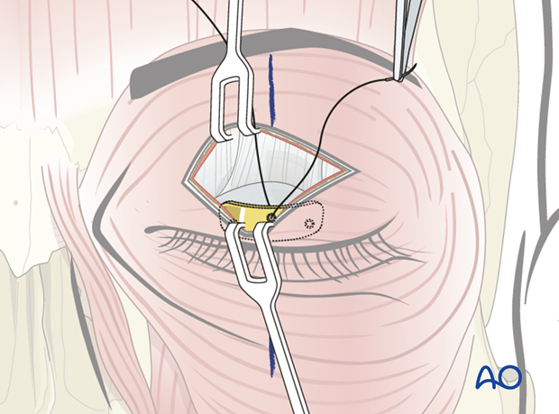
A 1-2 cm incision is centered over the mid pupillary line.
The cut is extended deep to the orbicularis muscle, but not beyond the levator.
Blunt dissection is performed to identify the tarsal plate.
Pocket preparation
A pocket for the placement of the weight is dissected.
This should be only slightly larger than the final implant, to allow insertion but preventing movement of the plate once in position.
Implant positioning
The weight should be centered over the mid pupillary line.
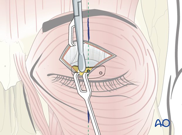
To increase the efficiency of closure the weight can be placed on the tarsal plate along the ciliary margin.
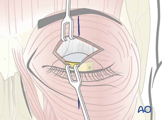
To reduce the postoperative visibility of the implant, the weight can also be placed higher, near the superior edge of the tarsal plate.
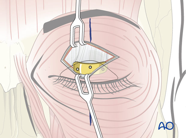
The weight is positioned inside the dedicated pocket in the desired position.
The implant is often sutured to the tarsus with a permanent or resorbable suture, to reduce the risk of movement.
Closure
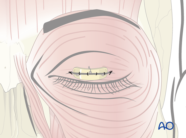
Once the weight is in position the skin incision is closed.
4. Case example: combined treatments
Patient with irreversible facial paralysis of the right side presenting with a painful, dry eye due to loss of eyelid closure, lower lid ectropion, and brow ptosis.
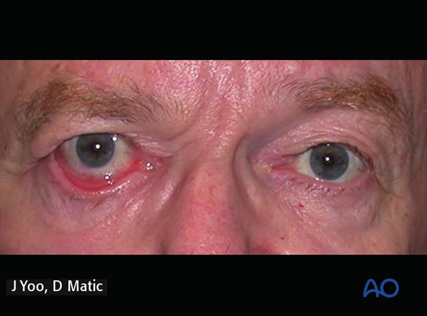
8 years postoperative following gold weight implant, direct brow lift, and transnasal lower lid tendon suspension.
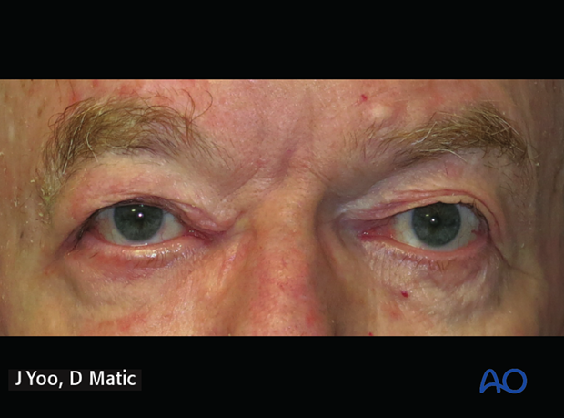
Video of this patient at 8 years follow-up
5. Aftercare
Lubrication
It is important to maintain lubrication and protection of the eye.
Lubrication consists of natural tear substitutes during the day and ointment based lubricants during sleep, often together with taping the eyelid closed.
Protection
Physical protection through eyewear may be necessary depending on occupation and environmental conditions.


