Lower eyelid - Tendon sling
1. Introduction
Tearing in the paralyzed eyelid
There are several reasons for excessive tearing that occurs in patients with eyelid paralysis:
- Exposure. As the cornea dries, this leads to more tear production.
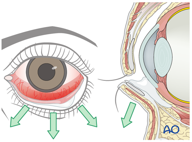
- Loss of lower eyelid support, secondary to flaccidity of the orbicularis oculi. As the lower lid falls away from the globe, the punctum and canalicular system work less efficiently to drain the tear film.
- The muscle driven pumping of the lacrimal sac cannot occur, which further increases tearing.
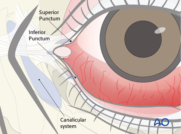
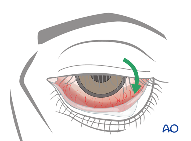
Closure of the upper eyelid reduces symptoms of exposure, but does not address the causes related to the drainage system. Therefore, improving lower eyelid position also needs to be considered to further reduce tearing.
Caution should be taken in performing a simultaneous upper lid procedure, as the requirement for upper lid closure may change following lower lid tightening and suspension.
The lower lid tendon suspension will improve upper lid closure even in the paralyzed eye by increasing the tension on the upper eyelid orbicularis oculi muscle.
Further considerations
The procedure of lower eyelid tendon sling is performed under general anesthetic.
2. Approach
Access to the medial orbital wall
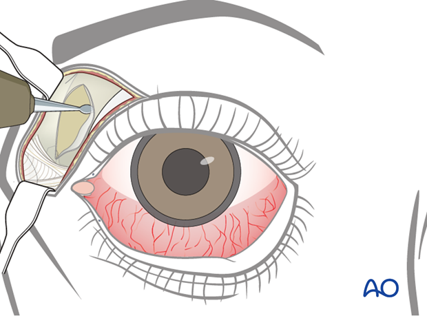
A modified blepharoplasty incision (15-20 mm) is made within the medial aspect of the supratarsal fold down through the orbicularis muscle.
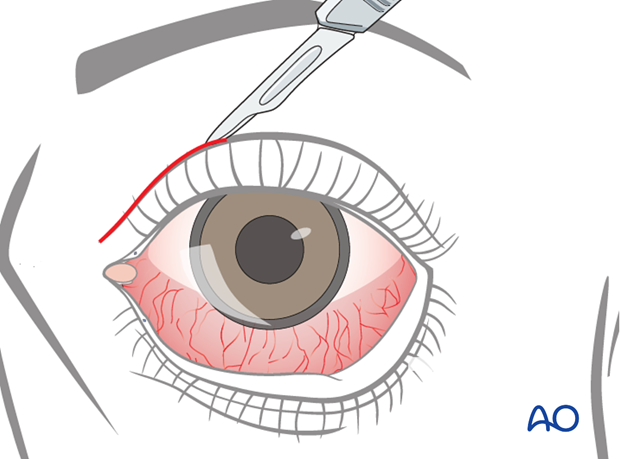
Medial traction is applied and an incision is made through the periosteum of the medial orbital wall.

Elevation of the periosteum of the medial wall
A periosteal elevator is used to elevate the periosteum of the medial wall of the nasofrontal junction and of the supraorbital rim.
Access to the lateral orbital wall
A lateral canthotomy or a cantholysis is performed.

Option: lateral tarsal strip
In some cases when the lower lid is also lax, lateral tarsal strip procedure can be performed in conjunction, which allows shortening of the lid by 5-8 mm.
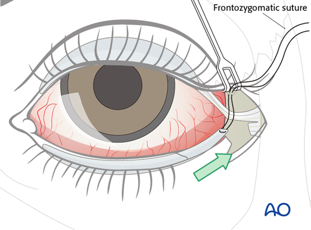
Elevation of the periosteum of the lateral wall
The periosteum of the lateral orbital rim is incised and elevated, exposing the lateral orbital rim and the internal lateral orbit.

3. Technique
Tendon harvest
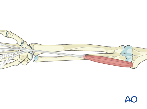
The most common tendon used is the palmaris longus.
In case of lack of this tendon on clinical examination, other options include plantaris or long extensors of the toes (3rd or 4th).
The selected tendon is harvested in full length.
Tendon preparation
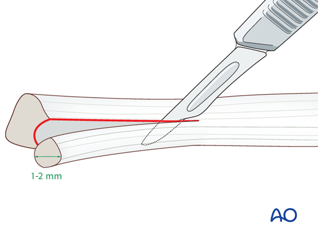
Once harvested, the tendon is split using a scalpel into a 1-2 mm thick strip.
Tendon sling positioning
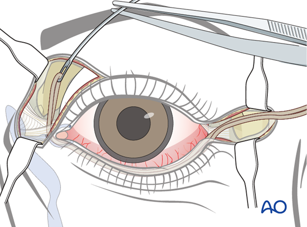
The harvested mini tendon is threaded along the ciliary margin of the lower lid using a Keith needle.
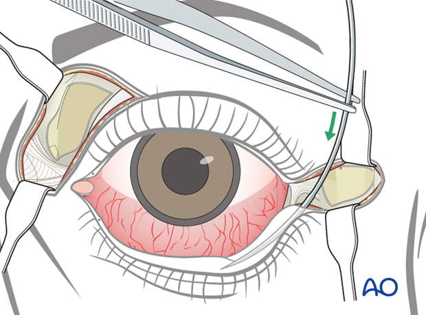
The needle can be pre-bent to better follow the gentle curve of the eyelid.
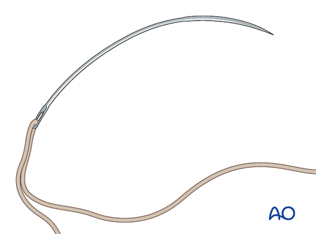
The needle is brought out of the skin just lateral to the inferior punctum.
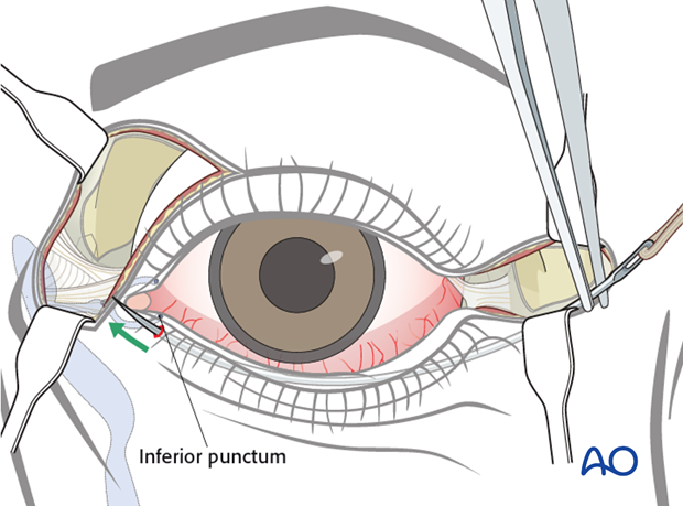
The needle is reinserted through the same puncture site, allowing a change in direction.
It is passed either through the medial canthal tendon, or deep to it avoiding the canalicular system.
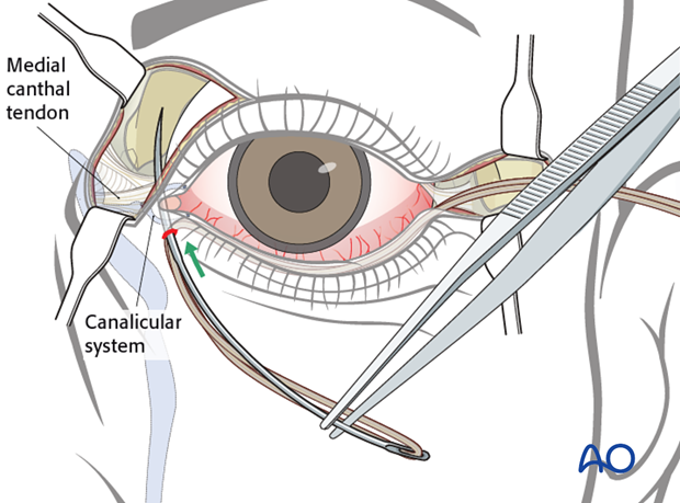
The needle exits subperiosteally and out through the upper eyelid incision, allowing the tendon to be threaded along the entire length of the lower eyelid.
Lateral fixation of the tendon sling
A 2.0 mm drill is used to drill a hole through the lateral orbital wall just below the frontozygomatic suture. The hole within the lateral orbit is placed 8-10 mm posterior to the orbital rim.
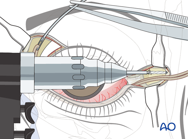
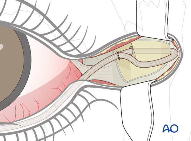
Laterally, the tendon is passed through the hole previously drilled in the lateral wall from medial to lateral. The tendon is looped and secured to itself using a 3-0 nonresorbable suture.
Medial tendon fastening
Medially, the end of the tendon is oversewn using a 3-0 nonresorbable suture to allow for sufficient purchase of the fixation wire.
The suture is passed multiple times through the tendon to sufficiently grab the tissue of the tendon.
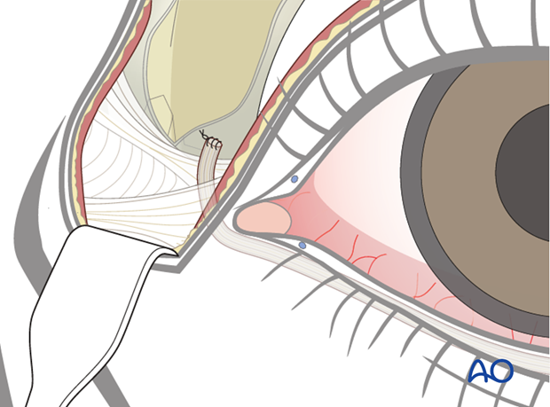
A 28 or 30 gauge stainless steel wire is passed through the suture, holding the tendon indirectly without risking pull through.
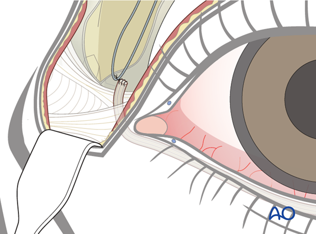
Medial plate anatomical adaptation
A plate is adapted to the anatomy of the anterior superior part of the medial orbital rim and medial wall.
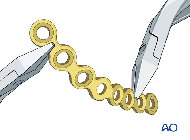
Medial plate positioning
The plate is applied on the anterior superior part of the medial orbital rim.
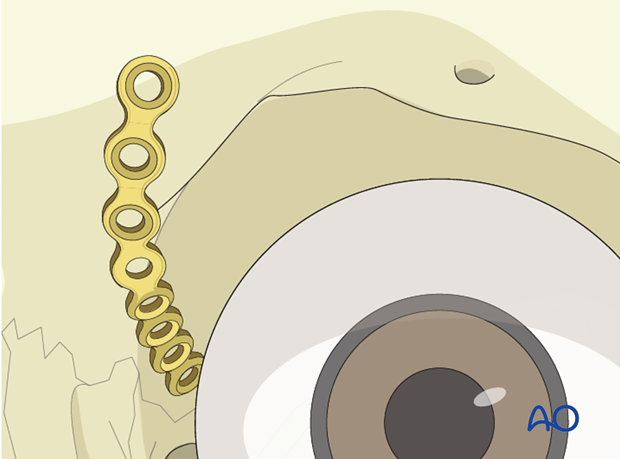
Medial fixation of the tendon sling
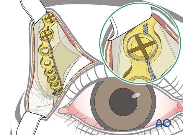
The tendon is placed superior and posterior to the lacrimal crest by passing the holding wire through the most posterior hole of the plate.
The tendon sling, once positioned, tightens the medial canthal tendon, the medial lower eyelid, and addresses the medial ectropion.

Tendon final tightening
Final tightening of the tendon sling is now performed.
Once satisfactory lower lid position and tension has been achieved, the wire can be twisted and tightened over one of the screws used to fix the plate or on a separate screw.
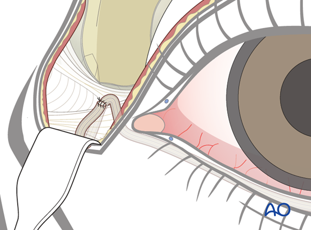
Alternative 1: tendon wrapped around the medial canthus
The tendon is wrapped around the medial canthus and sutured to it, with no bone fixation.
Advantages
- Technically easier
- Less morbidity
Disadvantage
- Does not adequately address medial lid and canthal laxity, often resulting in ongoing medial exposure of the globe
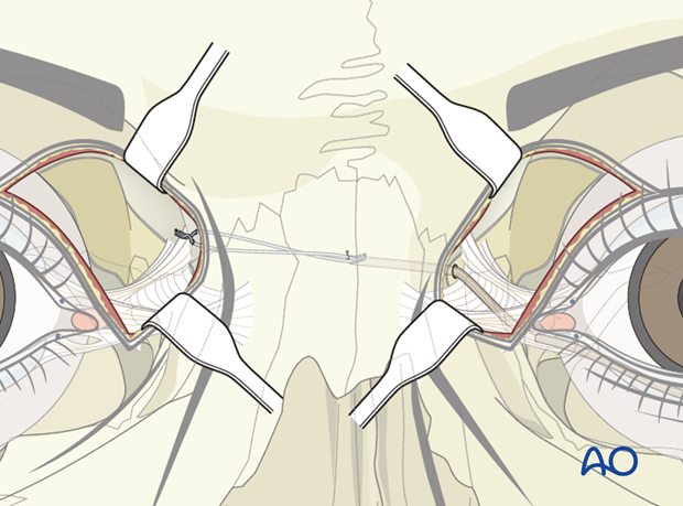
Alternative 2: transnasal tendon suspension
The medial tendon is secured trans nasally without the use of a plate.
Advantages
- May offer longer term stability of the tendon medially
- Reduced risk of the wire, suture, or tendon tearing on the medial orbital wall plate
Disadvantages
- Additional incision on the contralateral side
- Technically more challenging
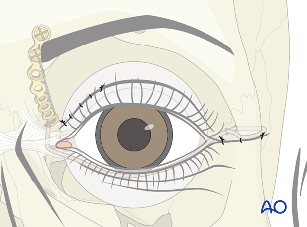
Closure
If a lateral strip canthopexy is planned, this is now completed.
Otherwise a layered closure of the cantholysis is completed.
4. Case example: combined treatments
Patient with irreversible facial paralysis of the right side presenting with a painful, dry eye due to loss of eyelid closure, lower lid ectropion, and brow ptosis.
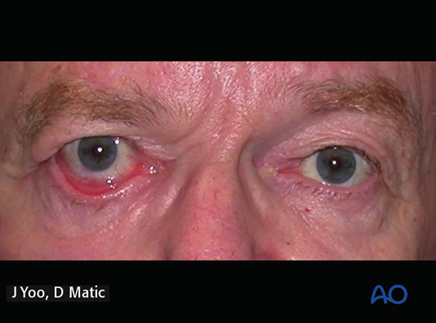
8 years postoperative following gold weight implant, direct brow lift, and transnasal lower lid tendon suspension.
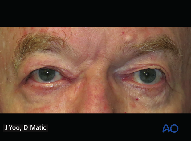
Video of this patient at 8 years follow-up
5. Aftercare
Antibiotic drops are provided 3-5 days postoperatively.
Lubrication
It is important to maintain lubrication and protection of the eye.
Lubrication consists of natural tear substitutes during the day and ointment based lubricants during sleep, often together with taping the eyelid closed.
Protection
Physical protection through eyewear may be necessary depending on occupation and environmental conditions.


