Open reduction internal fixation
1. General considerations
Due to the growth potential, the presence of tooth buds, and the rapid healing, pediatric surgical NOE fractures have special considerations:
- Small monocortical screws should be used to avoid injury to the developing tooth buds.
- Non-bioresorbable hardware should be removed once the fracture has healed.
NOE fractures in children, although rare in occurrence, require similar treatment as done with adults.
2. Case example
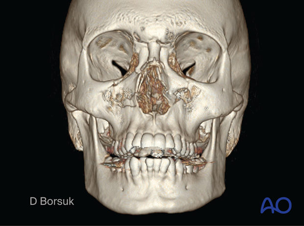
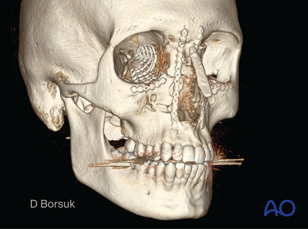
Frontal view CT scan of a 15-year-old with a right type II NOE fracture and left type I NOE fracture.
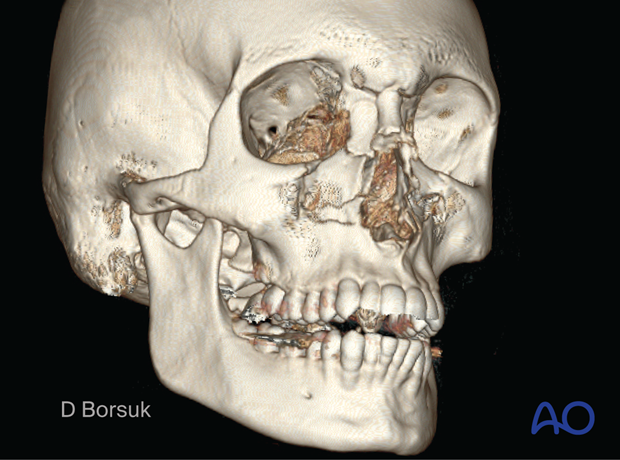
Oblique view of the same fracture.
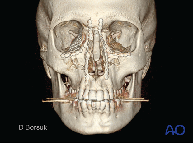
Postoperative frontal CT scan of the same patient. ORIF was done on the medial maxillary nasal frontal vertical buttress bilaterally, the inferior orbital rim and right internal orbit.
The nose was reduced, and additional primary bone graft was used for contour and projection.
Oblique view of the same patient.
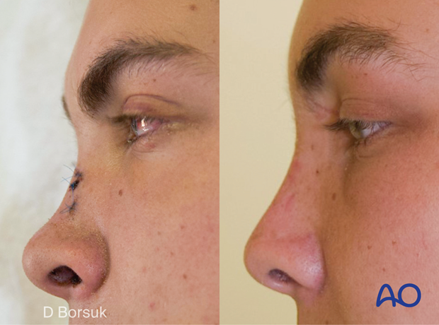
The left image shows preoperative lateral view displaying a collapsed upper central midface with a saddle nose deformity.
The right image displays restored projection of the nose and the upper central midface.
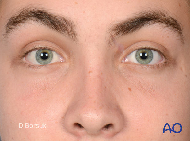
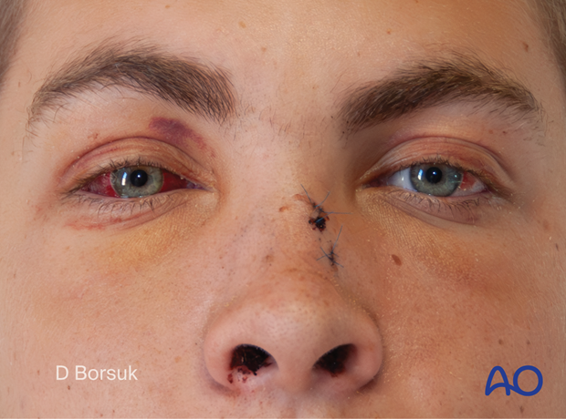
Frontal view of the preoperative bilateral NOE fracture displaying increased intercanthal distance and upper central facial collapse. Note the right globe enophthalmos.
Frontal postoperative view displaying restored upper central facial projection and restored intercanthal distance, medial canthal and globe positioning.