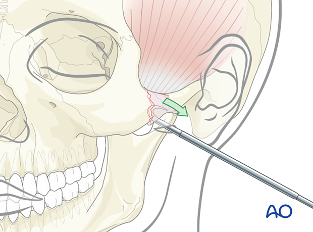
Indirect approaches to the zygomatic arch (temporal and transoral approaches)
1. General considerations
Proper alignment of the zygomatic arch facilitates the definition of the AP projection of the face. The width at the zygomatic arch defines the maximal width of the face.
The only direct exposure to the zygomatic arch is through a coronal incision. The coronal incision allows for excellent exposure of the zygomatic arch and reduction and fixation of comminuted fractures. However, the coronal incision results in significant scar alopecia and can also be associated with complications such as, temporal hollowing and possible risk of injury to the temporal branch of the facial nerve. In many instances, the negative sequelae of the coronal approach are worse than the benefit achieved from direct exposure of the arch. As a result, many surgeons prefer indirect exposures for the reduction of the zygomatic arch.
Indirect approaches
Common indirect approaches for reduction of the zygomatic arch include:
- Temporal (Gillies) approach (1)
- Transoral (Keen) approach (a lateral maxillary vestibular incision), (2)
- Percutaneous hook approach
An impact to the lateral side of the face can sometimes result in an isolated zygomatic arch fracture, where the zygomatic complex itself remains nondisplaced. In such cases, all three approaches can be considered to reduce the zygomatic arch.
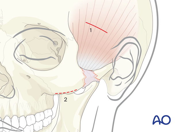
2. Temporal (Gillies) approach: Skin incision
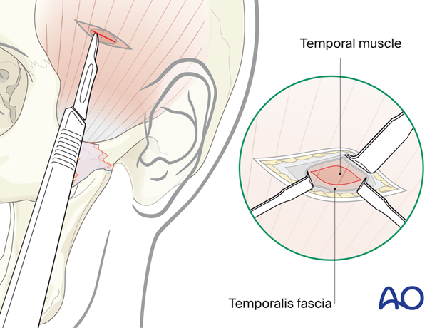
The Gillies technique describes a temporal incision (2 cm in length), made 2.5 cm superior and anterior to the helix, within the hairline.
After the injection of a local anesthetic with vasoconstrictor in the area of the proposed incision, a #15 blade is utilized to perform the incision. Care is taken to avoid the superficial temporal artery.
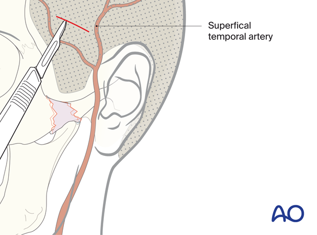
Temporal (Gillies) approach: Deep dissection
The dissection continues through the subcutaneous tissue and superficial temporal fascia down to the deep temporal fascia.
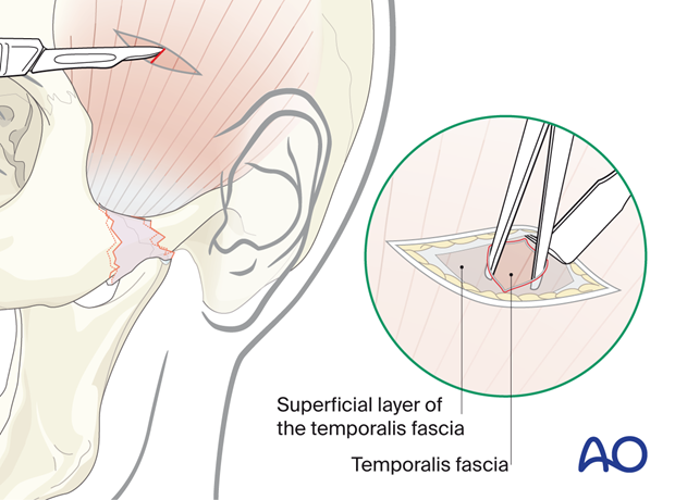
This fascia is incised to expose the temporalis muscle. The principal landmark is to visualize the fibers of the temporalis muscle.
Temporal (Gillies) approach: Exposure
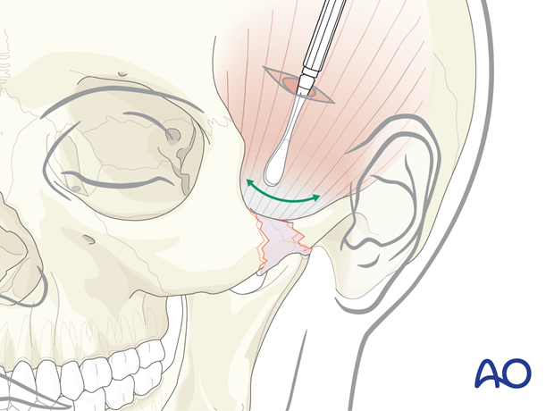
A freer elevator is inserted deep to the temporalis fascia and superficial to the temporalis muscle. The instrument is advanced using a back-and-forth motion until it is medial to the depressed zygomatic arch.
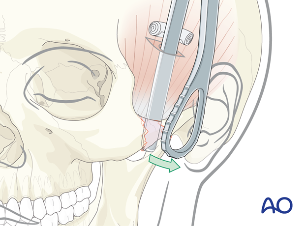
A Rowe zygomatic elevator is inserted just deep to the depressed zygomatic arch, and an outward force is applied.
Great care should be taken not to fulcrum off the squamous portion of the temporal bone.
Temporal (Gillies) approach: Wound closure
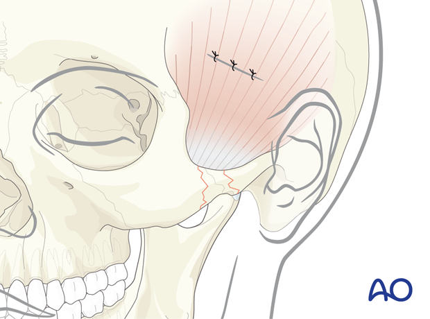
Closure is performed according to the preference of the surgeon.
3. Transoral (Keen) approach: Lateral maxillary vestibular incision
The transoral (Keen) approach provides indirect access to the zygomatic arch.
It allows for an intraoral incision and, therefore, avoids the risk of scar alopecia that can result from a temporal (Gillies) approach.
A 1 cm lateral maxillary vestibular incision (upper gingival buccal incision) is made with a scalpel or a cautery device just at the base of the zygomaticomaxillary buttress. The incision is made through mucosa only and blunt dissection is performed with a periosteal elevator behind the zygomaticomaxillary buttress proceeding to behind the zygomatic body and medial to the fractured zygomatic arch.
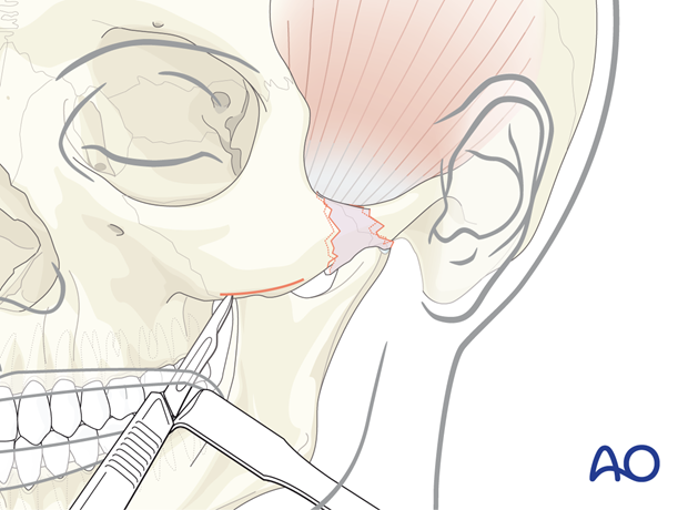
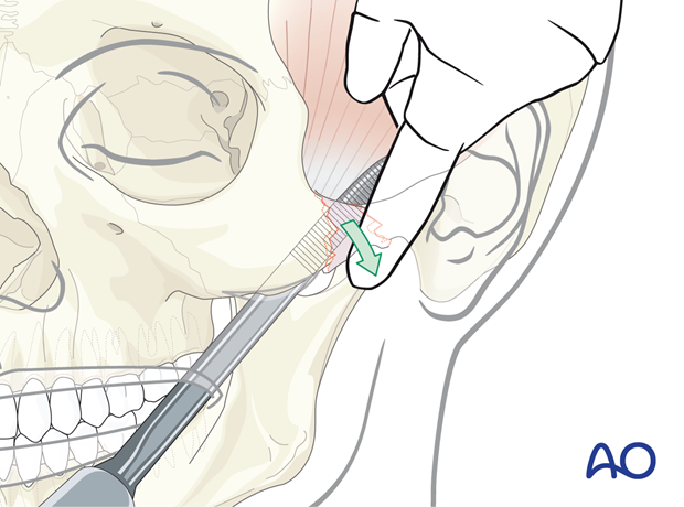
Transoral (Keen) approach: Exposure
Because of the direct proximity of the incision to the arch, an instrument can easily be placed deep to the fractured arch to allow its elevation. The arch can be palpated, elevated, and confirmed with a digital palpation.
4. Percutaneous hook approach
A percutaneous hook is placed through a small incision in the skin.
Poswillo draws two intersection lines on the face to determine the proper location for applying the bone hook. The first line is vertical, dropped down from the lateral cantus of the eye (involved fracture side).
The second line is horizontal, drawn laterally from the ala of the nose.
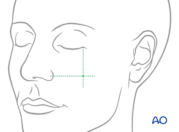
The percutaneous bone hook is placed through the skin around the depressed zygomatic fracture segments and pulled laterally.
