Combination of inferior and medial transconjunctival
1. Principles
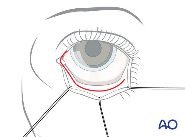
The pre-/transcaruncular approach can be combined with an inferior fornix transconjunctival approach. This combination allows exposure of the entire medial wall and the orbital floor.
The inferior fornix transconjunctival incision is extended mediosuperiorly with the incision running just anteriorly or directly through the caruncle.
The reverse sequence beginning with the pre-/transcaruncular incision and continuing in the lower fornix is very practical, too.
In both arrangements the medial orbit is dissected first. Read about the transconjunctival and the transcaruncular approach.
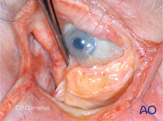
Inside the periorbita the medial pocket is extended anteroinferiorly around the lacrimal sac (nasolacrimal canal) and the soft tissues are spread until the inferior oblique muscle is encountered as shown in this anatomic series.
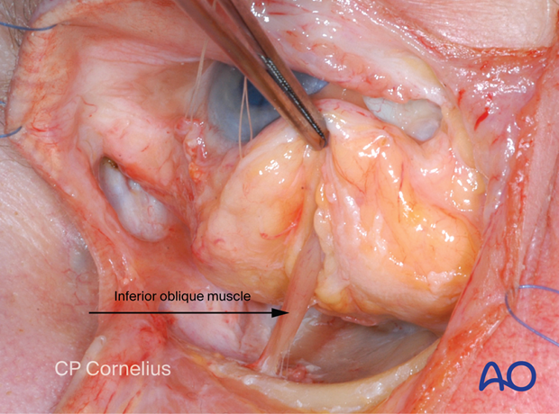
If a retroseptal route was chosen for the inferior fornix transconjunctival approach, the muscle insertion is horizontally transected leaving a small muscle cuff attached to the bone.
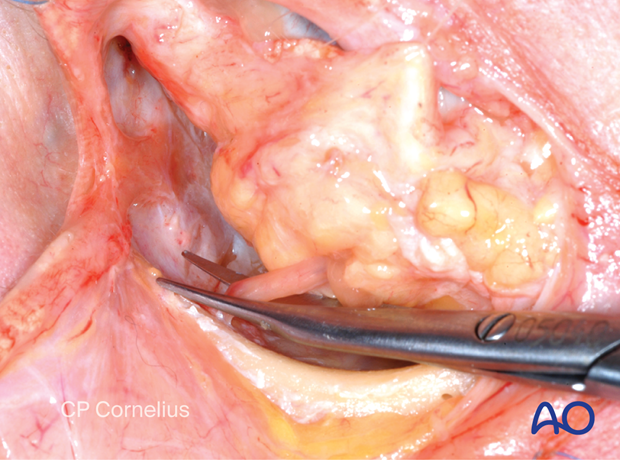
The periorbital incision between the medial pocket and posterior to the infraorbital rim are then connected. This allows for a joined exposure of the medial orbital wall and the orbital floor.
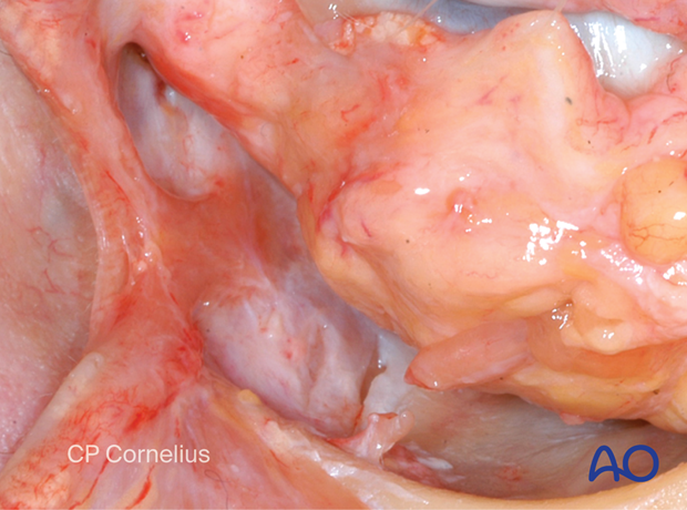
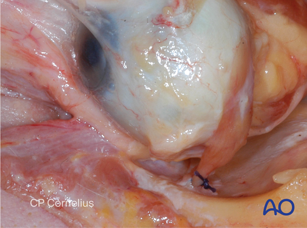
During closure, the inferior oblique muscle cuff is used to suture the belly of the muscle to its insertion.
If a preseptal route has been used for the inferior fornix transconjunctival approach the periorbita is incised medially to the inferior oblique muscle.
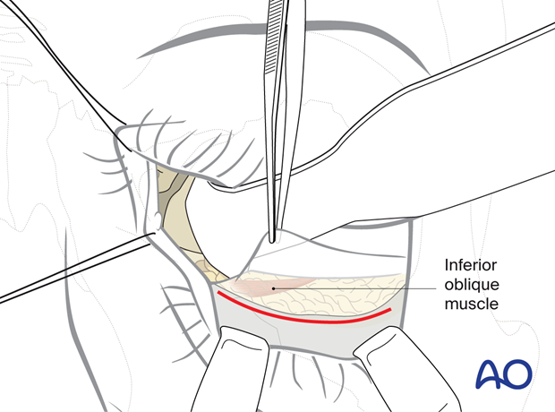
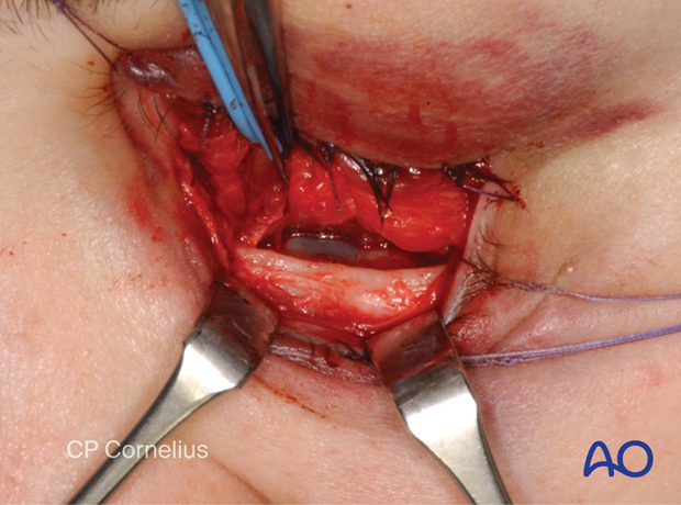
Then the muscle insertion is lifted together with the periorbital/subperiosteal dissection proceeding over the infraorbital rim into the orbital cavity.
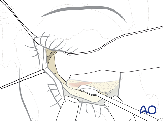
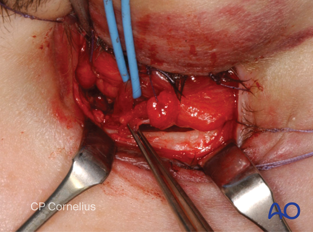
During closure the internal oblique muscle is reattached by simply suturing the periosteum.
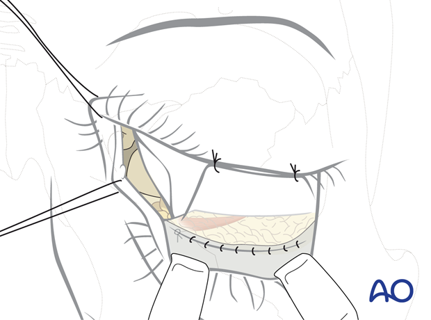
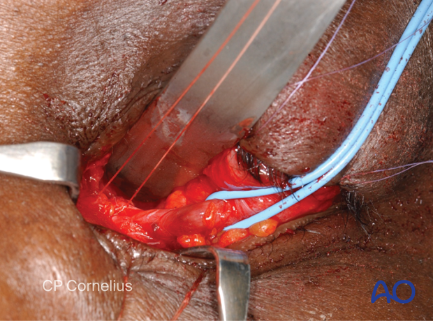
Clinical example showing the refixation technique for the internal oblique muscle during wound closure.
The muscle is marked with a blue rubber loop. Forceps at the periorbital incision edge.
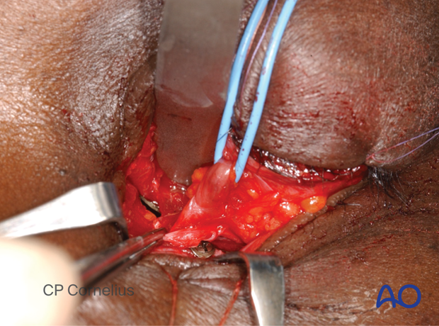
Suture guided for periorbital periosteal refixation.
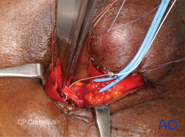
Suture tightened showing the reattachment of the muscle in the original anatomic position.
