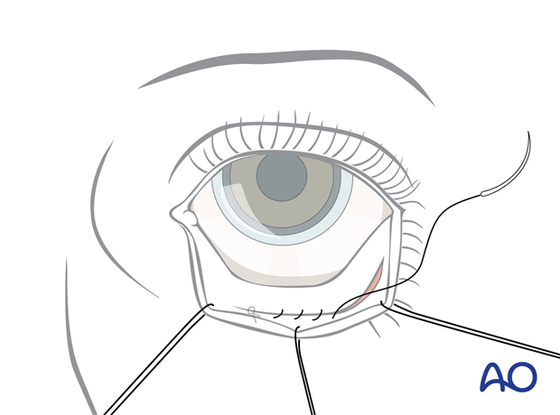
Inferior fornix transconjunctival approach
1. Principles
Surgical routes
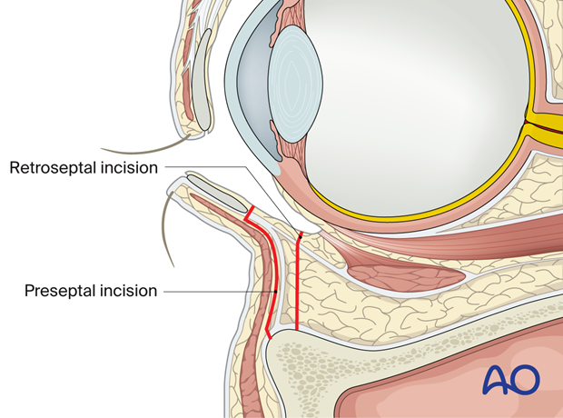
The typical inferior fornix transconjunctival approach can use two different routes to access the infraorbital rim:
- Retroseptal
- Preseptal
These two approaches vary in relation to the orbital septum on the pathway to the infraorbital rim.
Controversy exists on the advantages and disadvantages of these two surgical routes. We explain the retroseptal approach in more detail.
2. Retroseptal route
The retroseptal route enters directly into the fat compartments of the lower eyelids.
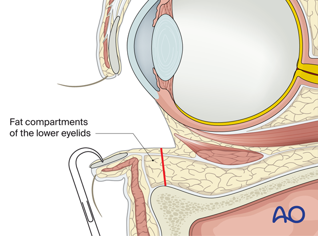
After vasoconstriction and insertion of a corneal shield two or three traction sutures are place through the lower eyelid.
After eversion of the lower eyelid, the position of the lower border of the tarsal plate is identified.
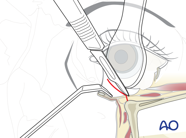
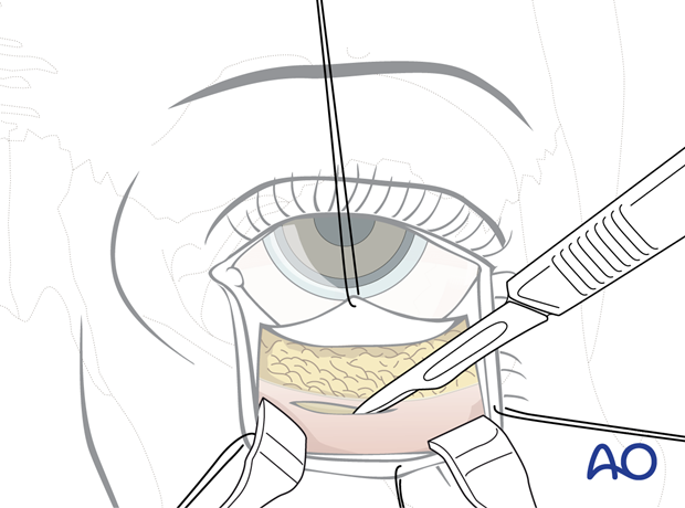
The incision is placed in the depth of the fornix. For the incision, scissors, scalpels, or electrocautery can be used.
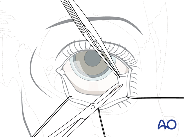
When using scissors, the conjunctiva is elevated laterally with a fine forceps and incised over a distance of about 8 mm including the lower-eyelid retractors.
3. Retroseptal incision and dissection
Starting from the initial transconjunctival incision, the conjunctiva and the lower-eyelid retractors are undermined towards the inner angle of the lids.
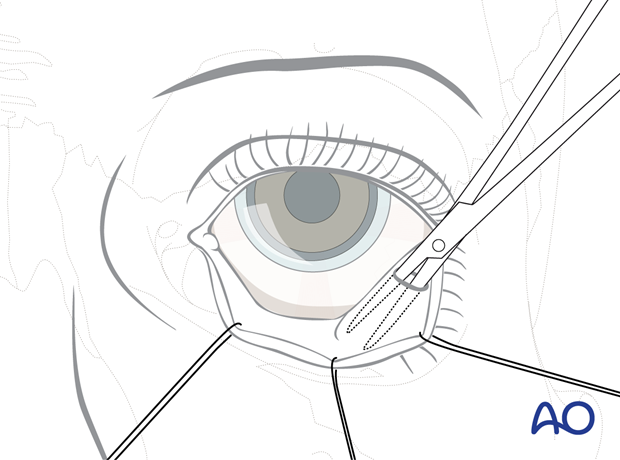
Then the incision line is extended from laterally to medially to expose the fat compartment behind the orbital septum.
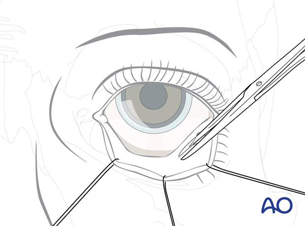
Alternative: scalpel or electrocautery incision
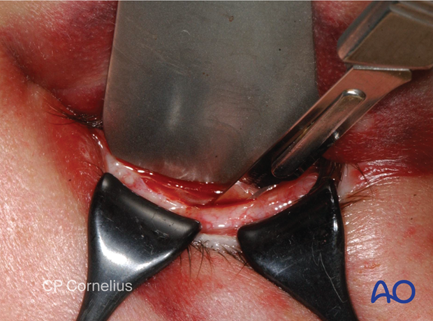
Instead of using scissors, the transsection of the conjunctiva and the lower-eyelid retractors can be carried out using a scalpel. Prior to this, the conjunctival tissues of the lower fornix are spanned over the infraorbital rim using a spatula and to small retractors.
4. Retroseptal incision of the periorbita
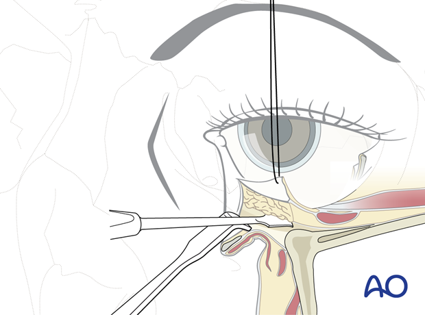
The lower lid is retracted downwards to expose the periorbita just behind the edge of the infraorbital rim.
(If it was decided to suture the posterior edge of the conjunctival flap as corneal protection, this is done now).
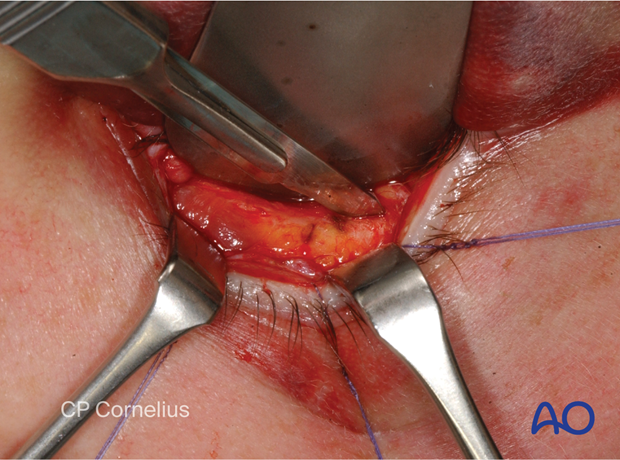
Next, the periorbita is incised parallel but just posterior to the infraorbital rim.
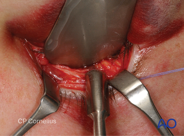
Once the infraorbital rim is exposed the periorbital dissection can be performed in the usual fashion.
Clinical photographs showing the incision and …
… dissection.
5. Retroseptal closure
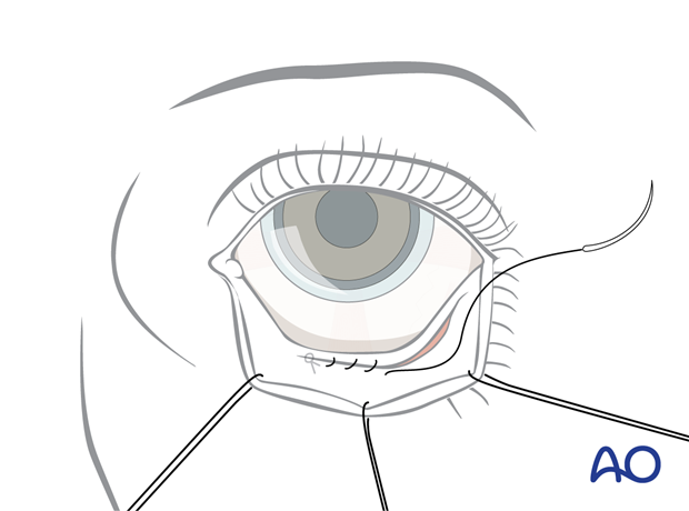
Closure of the periosteum or the periorbita respectively is not possible. The conjunctival incision is closed with a running 6-0 fast-absorbing suture. The suture ends may be buried or guided transcutaneously to bury the knots.
6. Preseptal route
The preseptal route requires entering the suborbicularis oculi/preseptal space above the fusion of the lower lid retractors and the orbital septum. This allows direct visualization of the septum.
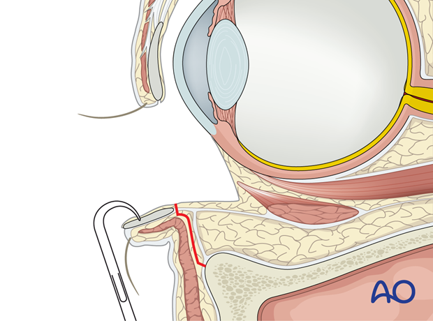
After vasoconstriction and insertion of a corneal shield, two or three traction sutures are place through the lower eyelid.
After eversion of the lower eyelid, the position of the lower border of the tarsal plate is identified.
The posterior edge of the conjunctiva is tented and secured with a stay suture and traction is applied to lift the conjunctiva while the lid margin is everted. The lower border of the tarsal plate is raised upward in this manner.
7. Preseptal incision and blunt dissection
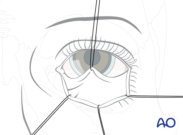
The incision is made directly below the lower tarsal border and the suborbicularis oculi/preseptal space is entered.
The incision is made using fine scissors starting laterally or a scalpel/electrocautery. Hemostasis is achieved with bipolar coagulation.
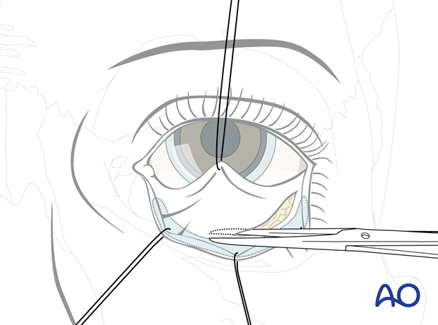
Blunt preseptal dissection is performed until the infraorbital rim is reached in the supraperiosteal plane.
At this point, the corneal shields are removed and the cephalic edge of the conjunctival flap is pulled up to be sutured to the upper lid for corneal protection.
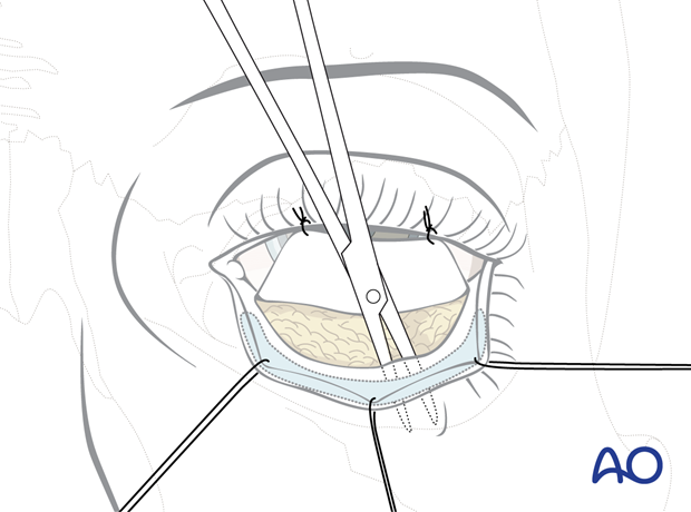
Once the infraorbital rim is exposed, the periorbital dissection can be performed in the usual fashion.
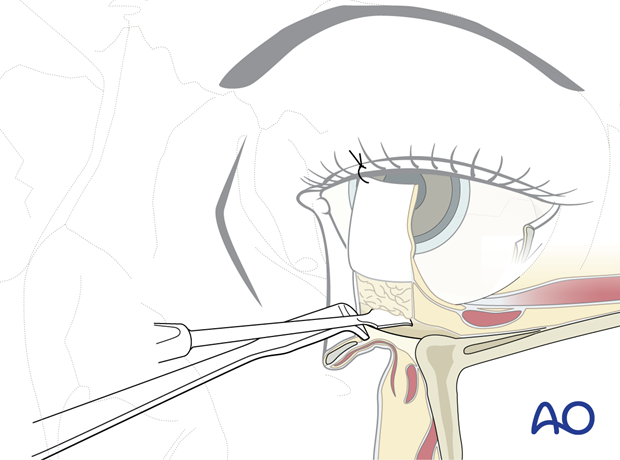
8. Preseptal closure
In the preseptal approach, the periosteum over the periorbital rim can be closed. The transconjunctival incision is closed using a 6-0 resorbable running suture with the suture ends either buried or guided through the skin externally to bury the knots.
An accurate alignment of the conjunctival flaps must be guaranteed.
